 Explore
 Validate
 Learn
Learn Western blot
Antibody data
- Antibody Data
- Antigen structure
- References [4]
- Comments [0]
- Validations
- Western blot [1]
- Flow cytometry [1]
- Other assay [3]
Submit
Validation data
Reference
Comment
Report error
- Product number
- PA5-25757 - Provider product page

- Provider
- Invitrogen Antibodies
- Product name
- PPAR gamma Polyclonal Antibody
- Antibody type
- Polyclonal
- Antigen
- Synthetic peptide
- Reactivity
- Human
- Host
- Rabbit
- Isotype
- IgG
- Vial size
- 400 µL
- Concentration
- 0.5 mg/mL
- Storage
- Store at 4°C short term. For long term storage, store at -20°C, avoiding freeze/thaw cycles.
Submitted references Erythropoietin Promotes Infection Resolution and Lowers Antibiotic Requirements in E. coli- and S. aureus-Initiated Infections.
IRF6 Regulates Alternative Activation by Suppressing PPARγ in Male Murine Macrophages.
Adipose tissue macrophages induce PPARγ-high FOXP3(+) regulatory T cells.
Developmental regulation of prostacyclin synthase and prostacyclin receptors in the ovine uterus and conceptus during the peri-implantation period.
Liang F, Guan H, Li W, Zhang X, Liu T, Liu Y, Mei J, Jiang C, Zhang F, Luo B, Zhang Z
Frontiers in immunology 2021;12:658715
Frontiers in immunology 2021;12:658715
IRF6 Regulates Alternative Activation by Suppressing PPARγ in Male Murine Macrophages.
Li C, Ying W, Huang Z, Brehm T, Morin A, Vella AT, Zhou B
Endocrinology 2017 Sep 1;158(9):2837-2847
Endocrinology 2017 Sep 1;158(9):2837-2847
Adipose tissue macrophages induce PPARγ-high FOXP3(+) regulatory T cells.
Onodera T, Fukuhara A, Jang MH, Shin J, Aoi K, Kikuta J, Otsuki M, Ishii M, Shimomura I
Scientific reports 2015 Nov 19;5:16801
Scientific reports 2015 Nov 19;5:16801
Developmental regulation of prostacyclin synthase and prostacyclin receptors in the ovine uterus and conceptus during the peri-implantation period.
Cammas L, Reinaud P, Bordas N, Dubois O, Germain G, Charpigny G
Reproduction (Cambridge, England) 2006 May;131(5):917-27
Reproduction (Cambridge, England) 2006 May;131(5):917-27
No comments: Submit comment
Supportive validation
- Submitted by
- Invitrogen Antibodies (provider)
- Main image
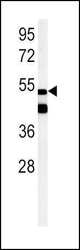
- Experimental details
- Western blot analysis in mouse heart tissue lysates (15 µg per lane) using a PPARG polyclonal antibody (Product # PA5-25757).

Supportive validation
- Submitted by
- Invitrogen Antibodies (provider)
- Main image

- Experimental details
- Flow cytometry analysis of HepG2 cells using a PPARG polyclonal antibody (Product # PA5-25757) (right) compared to a negative control cell (left) at a dilution of 1:10-50, followed by a FITC-conjugated goat anti-rabbit antibody

Supportive validation
- Submitted by
- Invitrogen Antibodies (provider)
- Main image

- Experimental details
- NULL

- Submitted by
- Invitrogen Antibodies (provider)
- Main image

- Experimental details
- Figure 3 NC-fed but not HFD mice-derived ATMs prompt PPARgamma-high Treg differentiation. ( A ) PPARgamma expression in Tregs induced by NC SPDCs and NC ATMs. ( B ) PPARgamma expression in Tregs induced by NC ATMs, HFD ATMs, and ob/ob ATMs. ( C ) Splenic T cells and adipose T cells isolated from NC and HFD mice were stained with CD25, FOXP3, and PPARgamma. The proportion of PPARgamma-high FOXP3 + Tregs was analyzed by FACS. ( D ) Numbers of PPARgamma-high Tregs in NC and ob/ob mice were counted by FACS. Data are representative of at least three independent experiments ( A - C ). Data are mean +- SEM. For ( A , B , D ): * P < 0.05, ** P < 0.01.

- Submitted by
- Invitrogen Antibodies (provider)
- Main image

- Experimental details
- Figure 4 EPO promotes macrophage clearance of E. coli through PPARgamma. (A, B) : Thioglycolate-elicited peritoneal macrophages from WT mice (A) or EPOR-cKO mice (B) were stimulated with rhEPO (20 IU/ml) or PBS in presence of heat deactivated E. coli (macrophage: E. coli = 1: 10) for 24 hrs and PPARgamma expression was evaluated by flow cytometry (n = 5). (C) : WT mice were inoculated with E. coli at 1 x 10 5 c.f.u. and peritoneal exudates of F4/80 + PPARgamma + macrophages were assayed by flow cytometry at indicated time points (n = 5). (D, E): WT (D) or EPOR-cKO (E) mice were inoculated with E. coli at 1 x 10 5 c.f.u. together with rhEPO (5,000 IU/kg) or PBS for 24 hrs and then peritoneal macrophage PPARgamma levels were measured (n = 5). (F-H) : Thioglycolate-elicited peritoneal macrophages were cultured with rhEPO (20 IU/ml, 24 hrs), RSG (10 muM, 48 hrs), GW9662 (10 muM, 48 hrs) or PBS and then incubated with fluorescently labeled E . coli for 30 min (macrophage: E. coli = 1: 10) for phagocytosis assay (n = 5). (I) : EPOR-cKO mice were pretreated with RSG (10 mg/kg, i.g.) or PBS for 5 days. On day 6, mice were inoculated with E. coli at 1 x 10 5 c.f.u. Intracellular E. coli levels in macrophages were analyzed at 24 hrs (n = 5). (J) : PPARgamma-C and PPARgamma-cKO mice were inoculated with E. coli at 10 5 c.f.u. together with rhEPO (5,000 IU/kg), RSG (pretreated by 10 mg/kg, i.g. for 5 days) or PBS for 24 hrs. Intracellular E. coli levels in macrophages were analyzed at 24
