Explore
 Validate
 Learn
Learn Western blot
Antibody data
- Antibody Data
- Antigen structure
- References [3]
- Comments [0]
- Validations
- Western blot [3]
- Immunocytochemistry [1]
- Immunoprecipitation [1]
- Flow cytometry [1]
Submit
Validation data
Reference
Comment
Report error
- Product number
- AF1584 - Provider product page

- Provider
- R&D Systems
- Product name
- Human/Mouse STAT5b Antibody
- Antibody type
- Polyclonal
- Description
- Antigen Affinity-purified. Detects human and mouse STAT5b.
- Reactivity
- Human, Mouse
- Host
- Rabbit
- Conjugate
- Unconjugated
- Isotype
- IgG
- Vial size
- 50 ug
- Concentration
- LYOPH
- Storage
- Use a manual defrost freezer and avoid repeated freeze-thaw cycles. 12 months from date of receipt, -20 to -70 °C as supplied. 1 month, 2 to 8 °C under sterile conditions after reconstitution. 6 months, -20 to -70 °C under sterile conditions after reconstitution.
Submitted references Critical functions for STAT5 tetramers in the maturation and survival of natural killer cells.
BCR-ABL affects STAT5A and STAT5B differentially.
Rac1 and a GTPase-activating protein, MgcRacGAP, are required for nuclear translocation of STAT transcription factors.
Lin JX, Du N, Li P, Kazemian M, Gebregiorgis T, Spolski R, Leonard WJ
Nature communications 2017 Nov 6;8(1):1320
Nature communications 2017 Nov 6;8(1):1320
BCR-ABL affects STAT5A and STAT5B differentially.
Schaller-Schönitz M, Barzan D, Williamson AJ, Griffiths JR, Dallmann I, Battmer K, Ganser A, Whetton AD, Scherr M, Eder M
PloS one 2014;9(5):e97243
PloS one 2014;9(5):e97243
Rac1 and a GTPase-activating protein, MgcRacGAP, are required for nuclear translocation of STAT transcription factors.
Kawashima T, Bao YC, Nomura Y, Moon Y, Tonozuka Y, Minoshima Y, Hatori T, Tsuchiya A, Kiyono M, Nosaka T, Nakajima H, Williams DA, Kitamura T
The Journal of cell biology 2006 Dec 18;175(6):937-46
The Journal of cell biology 2006 Dec 18;175(6):937-46
No comments: Submit comment
Supportive validation
- Submitted by
- R&D Systems (provider)
- Main image
- Experimental details
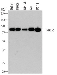
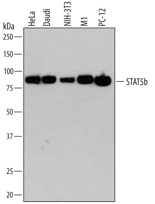
- Detection of Human, Mouse, and Rat STAT5b by Western Blot. Western blot shows lysates of HeLa human cervical epithelial carcinoma cell line, Daudi human Burkitt's lymphoma cell line, NIH-3T3 mouse embryonic fibroblast cell line, M1 mouse myeloid leukemia cell line, and PC-12 rat adrenal pheochromocytoma cell line. PVDF membrane was probed with 0.2 µg/mL of Rabbit Anti-Human/Mouse STAT5b Antigen Affinity-purified Polyclonal Antibody (Catalog # AF1584) followed by HRP-conjugated Anti-Rabbit IgG Secondary Antibody (Catalog # HAF008). A specific band was detected for STAT5b at approximately 90 kDa (as indicated). This experiment was conducted under reducing conditions and using Immunoblot Buffer Group 1.
- Submitted by
- R&D Systems (provider)
- Main image

- Experimental details
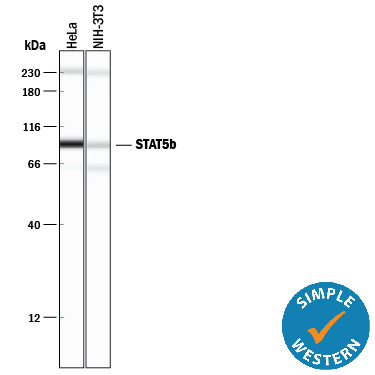
- Detection of Human and Mouse STAT5b by Simple WesternTM. Simple Western lane view shows lysates of HeLa human cervical epithelial carcinoma cell line and NIH-3T3 mouse embryonic fibroblast cell line, loaded at 0.2 mg/mL. A specific band was detected for STAT5b at approximately 92 kDa (as indicated) using 10 µg/mL of Rabbit Anti-Human/Mouse STAT5b Antigen Affinity-purified Polyclonal Antibody (Catalog # AF1584). This experiment was conducted under reducing conditions and using the 12-230 kDa separation system.Non-specific interaction with the 230 kDa Simple Western standard may be seen with this antibody.
- Submitted by
- R&D Systems (provider)
- Main image
- Experimental details
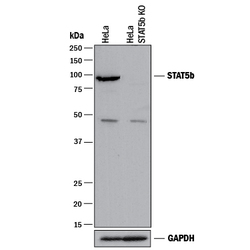
- Western Blot Shows Human STAT5b Specificity by Using Knockout Cell Line. Western blot shows lysates of HeLa human cervical epithelial carcinoma parental cell line and STAT5b knockout HeLa cell line (KO). PVDF membrane was probed with 0.2 µg/mL of Rabbit Anti-Human/Mouse STAT5b Antigen Affinity-purified Polyclonal Antibody (Catalog # AF1584) followed by HRP-conjugated Anti-Rabbit IgG Secondary Antibody (Catalog # HAF008). A specific band was detected for STAT5b at approximately 90 kDa (as indicated) in the parental HeLa cell line, but is not detectable in knockout HeLa cell line. GAPDH (Catalog # AF5718) is shown as a loading control. This experiment was conducted under reducing conditions and using Immunoblot Buffer Group 1.

Supportive validation
- Submitted by
- R&D Systems (provider)
- Main image

- Experimental details
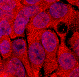
- STAT5b in HepG2 Human Cell Line. STAT5b was detected in immersion fixed HepG2 human hepatocellular carcinoma cell line using Rabbit Anti-Human/Mouse STAT5b Antigen Affinity-purified Polyclonal Antibody (Catalog # AF1584) at 5 µg/mL for 3 hours at room temperature. Cells were stained using the NorthernLights™ 557-conjugated Anti-Rabbit IgG Secondary Antibody (red; Catalog # NL004) and counterstained with DAPI(blue). Specific staining was localized to cytoplasm and nucleus. View our protocol for Fluorescent ICC Staining of Cells on Coverslips.
Supportive validation
- Submitted by
- R&D Systems (provider)
- Main image
- Experimental details
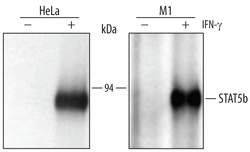
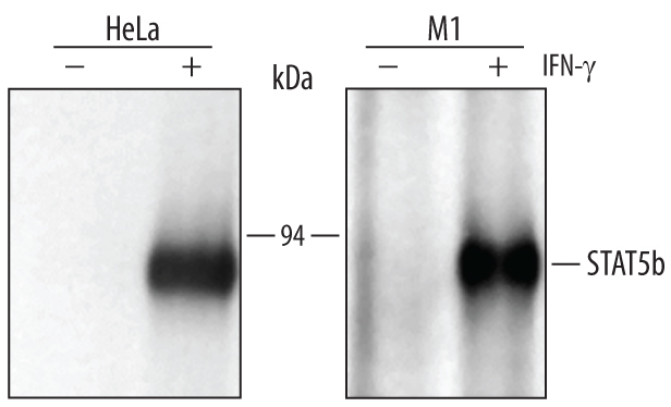
- Immunoprecipitation of Human and Mouse STAT5b. HeLa human cervical epithelial carcinoma and M1 mouse myeloid leukemia cell line were untreated (-) or activated (+) with 100 ng/mL Recombinant Human IFN-gamma (Catalog # 285-IF) for 15 minutes. STAT5b was immunoprecipitated from lysates of 5 x 10⁶ cells following incubation with 2 µg Rabbit Anti-Human/Mouse STAT5b Antigen Affinity-purified Polyclonal Antibody (Catalog # AF1584) for 1 hour at room temperature. STAT5b-antibody complexes were absorbed using Protein A sepharose (Invitrogen, Catalog # 10-1041). Immunoprecipitated STAT5b was detected by Western blot using 1 µg/mL Human Phospho-STAT5a/b (Y699) Antigen Affinity-purified Polyclonal Antibody (Catalog # AF4190). View our recommended buffer recipes for immunoprecipitation.
Supportive validation
- Submitted by
- R&D Systems (provider)
- Main image

- Experimental details
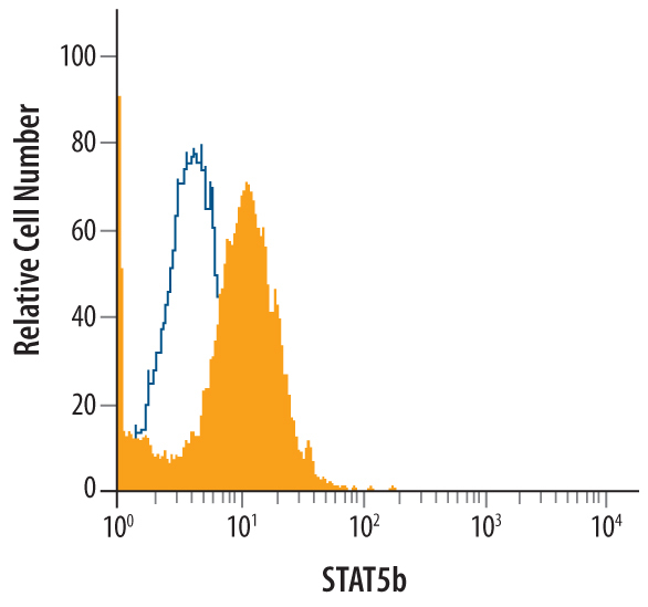
- Detection of STAT5b in Jurkat Human Cell Line by Flow Cytometry. Jurkat human acute T cell leukemia cell line was stained with Rabbit Anti-Human/Mouse STAT5b Antigen Affinity-purified Polyclonal Antibody (Catalog # AF1584, filled histogram) or control antibody (Catalog # AB-105-C, open histogram), followed by Allophycocyanin-conjugated Anti-Rabbit IgG Secondary Antibody (Catalog # F0111). To facilitate intracellular staining, cells were fixed with paraformaldehyde and permeabilized with methanol.