Explore
 Validate
 Learn
Learn Flow cytometry
 Other assay
Antibody data
- Antibody Data
- Antigen structure
- References [5]
- Comments [0]
- Validations
- Other assay [3]
Submit
Validation data
Reference
Comment
Report error
- Product number
- MHCD8605 - Provider product page

- Provider
- Invitrogen Antibodies
- Product name
- CD86 Monoclonal Antibody (BU63), APC
- Antibody type
- Monoclonal
- Antigen
- Other
- Description
- Allophycocyanin (APC) is a stable and highly soluble phycobiliprotein that provides maximal absorbance and fluorescence without susceptibility to internal or external fluorescence quenching, thus providing exceptional quantum yields and molar extinction coefficients.
- Reactivity
- Human
- Host
- Mouse
- Isotype
- IgG
- Antibody clone number
- BU63
- Vial size
- 500 µL
- Storage
- 4° C, store in dark
Submitted references Recombinant human IL-37 inhibited endometriosis development in a mouse model through increasing Th1/Th2 ratio by inducing the maturation of dendritic cells.
Expression of co-stimulatory molecules CD80 and CD86 is altered in CD14 + HLA-DR + monocytes from patients with Chagas disease following induction by Trypanosoma cruzi recombinant antigens.
Concurrent MEK and autophagy inhibition is required to restore cell death associated danger-signalling in Vemurafenib-resistant melanoma cells.
Antitumor immunity triggered by melphalan is potentiated by melanoma cell surface-associated calreticulin.
ROS-induced autophagy in cancer cells assists in evasion from determinants of immunogenic cell death.
Li L, Liao Z, Ye M, Jiang J
Reproductive biology and endocrinology : RB&E 2021 Aug 24;19(1):128
Reproductive biology and endocrinology : RB&E 2021 Aug 24;19(1):128
Expression of co-stimulatory molecules CD80 and CD86 is altered in CD14 + HLA-DR + monocytes from patients with Chagas disease following induction by Trypanosoma cruzi recombinant antigens.
Soares AK, Neves PA, Cavalcanti MD, Marinho SM, Oliveira W Júnior, Souza JR, Lorena VM, Gomes YM
Revista da Sociedade Brasileira de Medicina Tropical 2016 Sep-Oct;49(5):632-636
Revista da Sociedade Brasileira de Medicina Tropical 2016 Sep-Oct;49(5):632-636
Concurrent MEK and autophagy inhibition is required to restore cell death associated danger-signalling in Vemurafenib-resistant melanoma cells.
Martin S, Dudek-Perić AM, Maes H, Garg AD, Gabrysiak M, Demirsoy S, Swinnen JV, Agostinis P
Biochemical pharmacology 2015 Feb 1;93(3):290-304
Biochemical pharmacology 2015 Feb 1;93(3):290-304
Antitumor immunity triggered by melphalan is potentiated by melanoma cell surface-associated calreticulin.
Dudek-Perić AM, Ferreira GB, Muchowicz A, Wouters J, Prada N, Martin S, Kiviluoto S, Winiarska M, Boon L, Mathieu C, van den Oord J, Stas M, Gougeon ML, Golab J, Garg AD, Agostinis P
Cancer research 2015 Apr 15;75(8):1603-14
Cancer research 2015 Apr 15;75(8):1603-14
ROS-induced autophagy in cancer cells assists in evasion from determinants of immunogenic cell death.
Garg AD, Dudek AM, Ferreira GB, Verfaillie T, Vandenabeele P, Krysko DV, Mathieu C, Agostinis P
Autophagy 2013 Sep;9(9):1292-307
Autophagy 2013 Sep;9(9):1292-307
No comments: Submit comment
Supportive validation
- Submitted by
- Invitrogen Antibodies (provider)
- Main image

- Experimental details
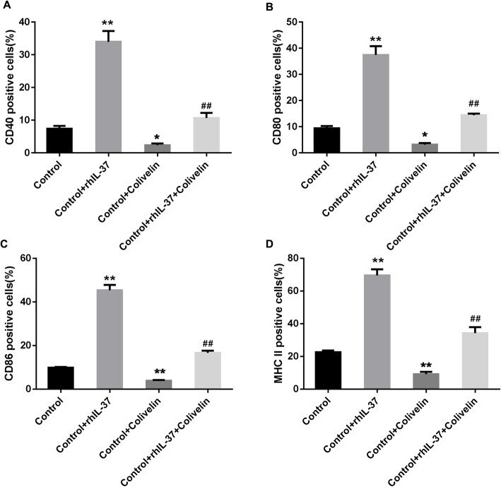
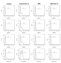
- Fig. 7 rhIL-37-induced the increasing of CD40, CD80, CD86, and MHC II in DCs was rescued by activation of STAT3. (A-D) The percentages of CD40-, CD80-, CD86-, and MHC II-positive DCs were determined using flow cytometry. N = 3. ** P < 0.01 compared with Control, and ## P < 0.01 compared with Control + rhIL-37
- Submitted by
- Invitrogen Antibodies (provider)
- Main image
- Experimental details
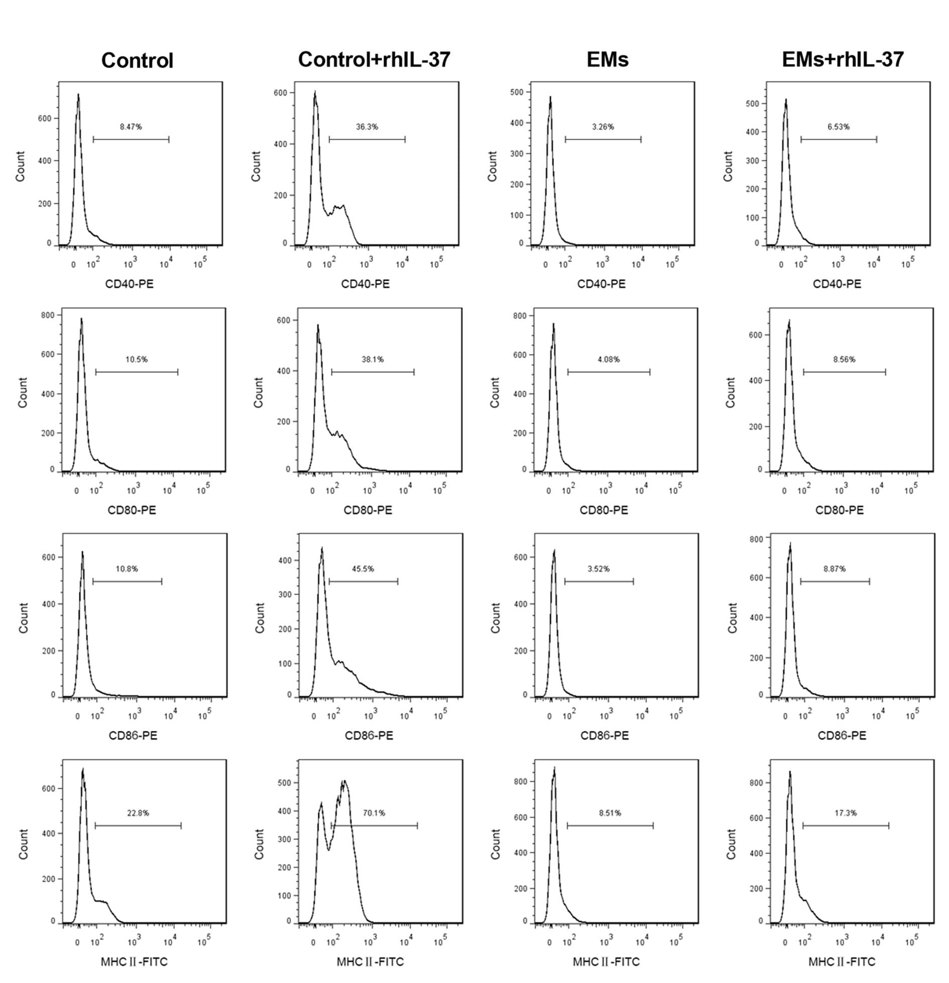
- Additional file 1: Supplementary figure 1 . Detection of the mature DCs percentage. At 24 hours after the last rhIL-37 administration, the percentages of CD40-, CD80-, CD86-, and MHC II-positive DCs in serum were determined using flow cytometry. N = 3.
- Submitted by
- Invitrogen Antibodies (provider)
- Main image
- Experimental details
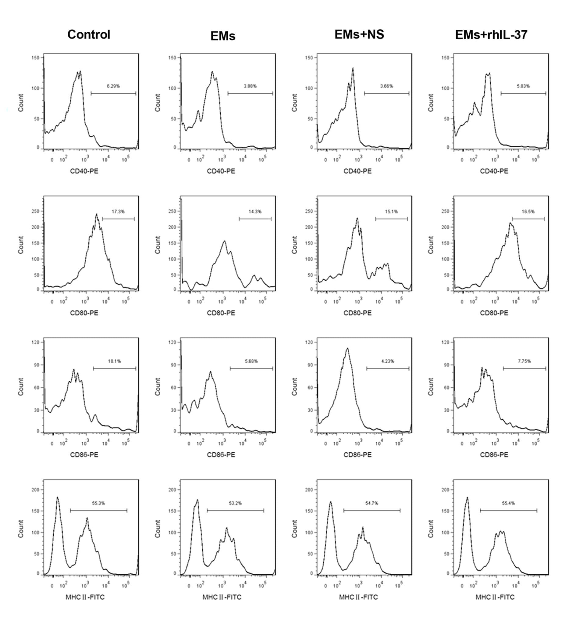
- Additional file 5: Supplementary figure 5 . Analysis of the maturation of DCs. DCs and EMs-DCs were separated, and were then treated with rhIL-37, and then the percentages of CD40-, CD80-, CD86-, and MHC II-positive DCs were determined using flow cytometry. N = 3.