Antibody data
- Antibody Data
- Antigen structure
- References [6]
- Comments [0]
- Validations
- Western blot [2]
- Immunohistochemistry [2]
- Blocking/Neutralizing [1]
Submit
Validation data
Reference
Comment
Report error
- Product number
- AF-141-NA - Provider product page

- Provider
- R&D Systems
- Product name
- Human B7-2/CD86 Antibody
- Antibody type
- Polyclonal
- Description
- Antigen Affinity-purified. Detects human B7-2/CD86 in direct ELISAs and Western blots. In direct ELISAs, less than 10% cross-reactivity with recombinant mouse B7-2 and recombinant rat B7-2 is observed.
- Reactivity
- Human
- Host
- Goat
- Conjugate
- Unconjugated
- Antigen sequence
P42081- Isotype
- IgG
- Vial size
- 200 ug
- Concentration
- LYOPH
- Storage
- Use a manual defrost freezer and avoid repeated freeze-thaw cycles. 12 months from date of receipt, -20 to -70 °C as supplied. 1 month, 2 to 8 °C under sterile conditions after reconstitution. 6 months, -20 to -70 °C under sterile conditions after reconstitution.
Submitted references Loss of 'homeostatic' microglia and patterns of their activation in active multiple sclerosis.
Tumor microenvironment contributes to Epstein-Barr virus anti-nuclear antigen-1 antibody production in nasopharyngeal carcinoma.
Regional immunity in melanoma: immunosuppressive changes precede nodal metastasis.
Modulation of T-cell activation by malignant melanoma initiating cells.
The soluble forms of CD28, CD86 and CTLA-4 constitute possible immunological markers in patients with abdominal aortic aneurysm.
Human plasma contains a soluble form of CD86 which is present at elevated levels in some leukaemia patients.
Zrzavy T, Hametner S, Wimmer I, Butovsky O, Weiner HL, Lassmann H
Brain : a journal of neurology 2017 Jul 1;140(7):1900-1913
Brain : a journal of neurology 2017 Jul 1;140(7):1900-1913
Tumor microenvironment contributes to Epstein-Barr virus anti-nuclear antigen-1 antibody production in nasopharyngeal carcinoma.
Ai P, Li Z, Jiang Y, Song C, Zhang L, Hu H, Wang T
Oncology letters 2017 Aug;14(2):2458-2462
Oncology letters 2017 Aug;14(2):2458-2462
Regional immunity in melanoma: immunosuppressive changes precede nodal metastasis.
Mansfield AS, Holtan SG, Grotz TE, Allred JB, Jakub JW, Erickson LA, Markovic SN
Modern pathology : an official journal of the United States and Canadian Academy of Pathology, Inc 2011 Apr;24(4):487-94
Modern pathology : an official journal of the United States and Canadian Academy of Pathology, Inc 2011 Apr;24(4):487-94
Modulation of T-cell activation by malignant melanoma initiating cells.
Schatton T, Schütte U, Frank NY, Zhan Q, Hoerning A, Robles SC, Zhou J, Hodi FS, Spagnoli GC, Murphy GF, Frank MH
Cancer research 2010 Jan 15;70(2):697-708
Cancer research 2010 Jan 15;70(2):697-708
The soluble forms of CD28, CD86 and CTLA-4 constitute possible immunological markers in patients with abdominal aortic aneurysm.
Sakthivel P, Shively V, Kakoulidou M, Pearce W, Lefvert AK
Journal of internal medicine 2007 Apr;261(4):399-407
Journal of internal medicine 2007 Apr;261(4):399-407
Human plasma contains a soluble form of CD86 which is present at elevated levels in some leukaemia patients.
Hock BD, Patton WN, Budhia S, Mannari D, Roberts P, McKenzie JL
Leukemia 2002 May;16(5):865-73
Leukemia 2002 May;16(5):865-73
No comments: Submit comment
Supportive validation
- Submitted by
- R&D Systems (provider)
- Main image
- Experimental details
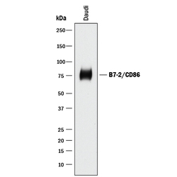
- Detection of Human B7-2/CD86 by Western Blot. Western blot shows lysates of Daudi human Burkitt's lymphoma cell line. PVDF membrane was probed with 0.5 µg/mL of Goat Anti-Human B7-2/CD86 Antigen Affinity-purified Polyclonal Antibody (Catalog # AF-141-NA) followed by HRP-conjugated Anti-Goat IgG Secondary Antibody (Catalog # HAF017). A specific band was detected for B7-2/CD86 at approximately 75 kDa (as indicated). This experiment was conducted under reducing conditions and using Immunoblot Buffer Group 1.
- Submitted by
- R&D Systems (provider)
- Main image
- Experimental details
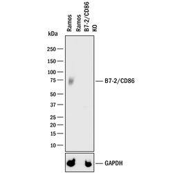
- Western Blot Shows Human B7-2/CD86 Specificity by Using Knockout Cell Line. Western blot shows lysates of Ramos human Burkitt's lymphoma parental cell line and B7-2/CD86 knockout Ramos cell line (KO). PVDF membrane was probed with 0.5 µg/mL of Goat Anti-Human B7-2/CD86 Antigen Affinity-purified Polyclonal Antibody (Catalog # AF-141-NA) followed by HRP-conjugated Anti-Goat IgG Secondary Antibody (Catalog # HAF017). A specific band was detected for B7-2/CD86 at approximately 74 kDa (as indicated) in the parental Ramos cell line, but is not detectable in knockout Ramos cell line. GAPDH (Catalog # AF5718) is shown as a loading control. This experiment was conducted under reducing conditions and using Immunoblot Buffer Group 1.
Supportive validation
- Submitted by
- R&D Systems (provider)
- Main image
- Experimental details
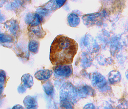
- B7-2/CD86 in Human Tonsil. B7-2/CD86 was detected in immersion fixed paraffin-embedded sections of human tonsil using Goat Anti-Human B7-2/CD86 Antigen Affinity-purified Polyclonal Antibody (Catalog # AF-141-NA) at 15 µg/mL overnight at 4 °C. Tissue was stained using the Anti-Goat HRP-DAB Cell & Tissue Staining Kit (brown; Catalog # CTS008) and counterstained with hematoxylin (blue). View our protocol for Chromogenic IHC Staining of immersion fixed paraffin-embedded Tissue Sections.
- Submitted by
- R&D Systems (provider)
- Main image
- Experimental details
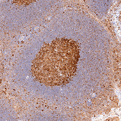
- B7-2/CD86 in Human Tonsil. B7-2/CD86 was detected in immersion fixed paraffin-embedded sections of human tonsil using Goat Anti-Human B7-2/CD86 Antigen Affinity-purified Polyclonal Antibody (Catalog # AF-141-NA) at 3 µg/mL for 1 hour at room temperature followed by incubation with the Anti-Goat IgG VisUCyte™ HRP Polymer Antibody (Catalog # VC004). Tissue was stained using DAB (brown) and counterstained with hematoxylin (blue). Specific staining was localized to lymphocytes. View our protocol for IHC Staining with VisUCyte HRP Polymer Detection Reagents.
Supportive validation
- Submitted by
- R&D Systems (provider)
- Main image
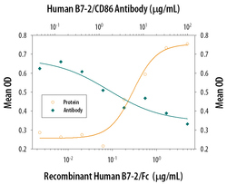
- Experimental details
- IL-2 secretion Induced by B7-2/CD86 and Neutralization by Human B7-2/CD86 Antibody. Recombinant Human B7-2/CD86 Fc Chimera (Catalog # 141-B2) co-stimulates IL-2 secretion in the Jurkat human acute T cell leukemia cell line in the presence of PHA in a dose-dependent manner (orange line), as measured by the Human IL-2 Quantikine ELISA Kit (Catalog # D2050). IL-2 secretion elicited by Recombinant Human B7-2/CD86 Fc Chimera (2 µg/mL) and PHA (10 µg/mL) is neutralized (green line) by increasing concentrations of Goat Anti-Human B7-2/CD86 Antigen Affinity-purified Polyclonal Antibody (Catalog # AF-141-NA). The ND50 is typically 0.25-1.25 µg/mL.
 Explore
 Validate
 Learn
Learn Western blot