Explore
 Validate
 Learn
Learn Western blot
Antibody data
- Antibody Data
- Antigen structure
- References [3]
- Comments [0]
- Validations
- Western blot [1]
Submit
Validation data
Reference
Comment
Report error
- Product number
- PAB9986 - Provider product page

- Provider
- Abnova Corporation
- Proper citation
- Abnova Corporation Cat#PAB9986, RRID:AB_1672192
- Product name
- CDC27 polyclonal antibody
- Antibody type
- Polyclonal
- Description
- Rabbit polyclonal antibody raised against synthetic peptide of CDC27.
- Storage
- Store at 4°C. For long term storage store at -20°C.Aliquot to avoid repeated freezing and thawing.
Submitted references Early mitotic degradation of the homeoprotein HOXC10 is potentially linked to cell cycle progression.
The dephosphorylated form of the anaphase-promoting complex protein Cdc27/Apc3 concentrates on kinetochores and chromosome arms in mitosis.
Mad2 transiently associates with an APC/p55Cdc complex during mitosis.
Gabellini D, Colaluca IN, Vodermaier HC, Biamonti G, Giacca M, Falaschi A, Riva S, Peverali FA
The EMBO journal 2003 Jul 15;22(14):3715-24
The EMBO journal 2003 Jul 15;22(14):3715-24
The dephosphorylated form of the anaphase-promoting complex protein Cdc27/Apc3 concentrates on kinetochores and chromosome arms in mitosis.
Topper LM, Campbell MS, Tugendreich S, Daum JR, Burke DJ, Hieter P, Gorbsky GJ
Cell cycle (Georgetown, Tex.) 2002 Jul-Aug;1(4):282-92
Cell cycle (Georgetown, Tex.) 2002 Jul-Aug;1(4):282-92
Mad2 transiently associates with an APC/p55Cdc complex during mitosis.
Wassmann K, Benezra R
Proceedings of the National Academy of Sciences of the United States of America 1998 Sep 15;95(19):11193-8
Proceedings of the National Academy of Sciences of the United States of America 1998 Sep 15;95(19):11193-8
No comments: Submit comment
Supportive validation
- Submitted by
- Abnova Corporation (provider)
- Main image

- Experimental details
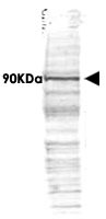
- Western blot using CDC27 polyclonal antibody (Cat # PAB9986) shows detection of a band ~90 KDa corresponding to human CDC27 (arrowhead).Approximately 35 ug of HeLa whole cell lysate was separated by SDS-PAGE and transferred onto nitrocellulose.After blocking the membrane was probed with the primary antibody diluted to 1.0 ug/mL for 2 h atroom temperature followed by washes and reaction with a 1 : 10,000 dilution of IRDye™800 conjugated Gt-a-Rabbit IgG [H&L] MX for 45 min at room temperature.IRDye™800 fluorescence image was captured using the Odyssey® Infrared Imaging System developedby LI-COR.IRDye is a trademark of LI-COR, Inc.