Explore
 Validate
 Learn
Learn Flow cytometry
Antibody data
- Antibody Data
- Antigen structure
- References [6]
- Comments [0]
- Validations
- Flow cytometry [1]
- Other assay [6]
Submit
Validation data
Reference
Comment
Report error
- Product number
- 12-1199-42 - Provider product page

- Provider
- Invitrogen Antibodies
- Product name
- CD119 (IFN gamma Receptor 1) Monoclonal Antibody (GIR-208 (GIR 208)), PE, eBioscience™
- Antibody type
- Monoclonal
- Antigen
- Other
- Description
- Description: The monoclonal antibody reacts with human CD119 also known as the IFN gamma receptor 1 (alpha chain). IFN gamma R1 is found in a complex with IFN gamma receptor beta and recognizes with high affinity IFN gamma. Expression is ubiquitous but low, although some cell types, such as monocytes express higher levels. The GIR-208 monoclonal antibody is blocked from binding when IFN gamma is bound to the receptor. Applications Reported: This GIR-208 (GIR 208) antibody has been reported for use in flow cytometric analysis. Applications Tested: This GIR-208 (GIR 208) antibody has been pre-titrated and tested by flow cytometric analysis of normal human peripheral blood cells. This can be used at 5 µL (0.5 µg) per test. A test is defined as the amount (µg) of antibody that will stain a cell sample in a final volume of 100 µL. Cell number should be determined empirically but can range from 10^5 to 10^8 cells/test. Excitation: 488-561 nm; Emission: 578 nm; Laser: Blue Laser, Green Laser, Yellow-Green Laser. Filtration: 0.2 µm post-manufacturing filtered.
- Reactivity
- Human
- Host
- Mouse
- Conjugate
- Yellow dye
- Isotype
- IgG
- Antibody clone number
- GIR-208 (GIR 208)
- Vial size
- 100 Tests
- Concentration
- 5 µL/Test
- Storage
- 4° C, store in dark, DO NOT FREEZE!
Submitted references Mechanistic and pharmacodynamic studies of DuoBody-CD3x5T4 in preclinical tumor models.
Human Lentiviral Gene Therapy Restores the Cellular Phenotype of Autosomal Recessive Complete IFN-γR1 Deficiency.
Patient iPSC-Derived Macrophages to Study Inborn Errors of the IFN-γ Responsive Pathway.
Mycobacterium tuberculosis replicates within necrotic human macrophages.
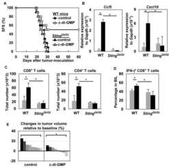
STING contributes to antiglioma immunity via triggering type I IFN signals in the tumor microenvironment.
Generation and characterization of monoclonal antibodies specific for the human IFN-gamma receptor.
Kemper K, Gielen E, Boross P, Houtkamp M, Plantinga TS, de Poot SA, Burm SM, Janmaat ML, Koopman LA, van den Brink EN, Rademaker R, Verzijl D, Engelberts PJ, Satijn D, Sasser AK, Breij EC
Life science alliance 2022 Nov;5(11)
Life science alliance 2022 Nov;5(11)
Human Lentiviral Gene Therapy Restores the Cellular Phenotype of Autosomal Recessive Complete IFN-γR1 Deficiency.
Hahn K, Pollmann L, Nowak J, Nguyen AHH, Haake K, Neehus AL, Waqas SFH, Pessler F, Baumann U, Hetzel M, Casanova JL, Schulz A, Bustamante J, Ackermann M, Lachmann N
Molecular therapy. Methods & clinical development 2020 Jun 12;17:785-795
Molecular therapy. Methods & clinical development 2020 Jun 12;17:785-795
Patient iPSC-Derived Macrophages to Study Inborn Errors of the IFN-γ Responsive Pathway.
Haake K, Neehus AL, Buchegger T, Kühnel MP, Blank P, Philipp F, Oleaga-Quintas C, Schulz A, Grimley M, Goethe R, Jonigk D, Kalinke U, Boisson-Dupuis S, Casanova JL, Bustamante J, Lachmann N
Cells 2020 Feb 19;9(2)
Cells 2020 Feb 19;9(2)
Mycobacterium tuberculosis replicates within necrotic human macrophages.
Lerner TR, Borel S, Greenwood DJ, Repnik U, Russell MR, Herbst S, Jones ML, Collinson LM, Griffiths G, Gutierrez MG
The Journal of cell biology 2017 Mar 6;216(3):583-594
The Journal of cell biology 2017 Mar 6;216(3):583-594
STING contributes to antiglioma immunity via triggering type I IFN signals in the tumor microenvironment.
Ohkuri T, Ghosh A, Kosaka A, Zhu J, Ikeura M, David M, Watkins SC, Sarkar SN, Okada H
Cancer immunology research 2014 Dec;2(12):1199-208
Cancer immunology research 2014 Dec;2(12):1199-208
Generation and characterization of monoclonal antibodies specific for the human IFN-gamma receptor.
Sheehan KC, Calderon J, Schreiber RD
Journal of immunology (Baltimore, Md. : 1950) 1988 Jun 15;140(12):4231-7
Journal of immunology (Baltimore, Md. : 1950) 1988 Jun 15;140(12):4231-7
No comments: Submit comment
Supportive validation
- Submitted by
- Invitrogen Antibodies (provider)
- Main image

- Experimental details
- Staining of normal human peripheral blood cells with Mouse IgG1 kappa Isotype Control PE (Product # 12-4714-81) (open histogram) or Anti-Human CD119 (IFN gamma Receptor 1) PE (filled histogram). Cells in the lymphocyte (left) or monocyte (right) gates were used for analysis.
- Conjugate
- Yellow dye
Supportive validation
- Submitted by
- Invitrogen Antibodies (provider)
- Main image
- Experimental details
- NULL
- Conjugate
- Yellow dye

- Submitted by
- Invitrogen Antibodies (provider)
- Main image
- Experimental details
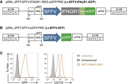
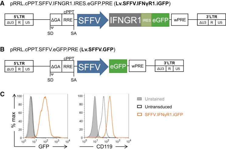
- Figure 1 Lentiviral Vector Design and Evaluation of Transgene Expression (A and B) Schematic overview of the third-generation SIN lentiviral vectors encoding (A) IFNGR1 cDNA coupled to a GFP reporter via an internal ribosomal entry side (IRES) and (B) a control vector encoding only for the GFP reporter. Transgene expression is driven by a spleen focus forming virus (SFFV) promoter in both constructs. (C) Flow cytometric analysis of GFP and IFN-gammaR1 (CD119) expression in untransduced and Lv.SFFV.IFNgammaR1.iGFP-transduced K562 cells (human myeloid cell line; gray filled: unstained untransduced cells, black: stained untransduced cells, orange: Lv.SFFV.IFNgammaR1.iGFP-transduced K562 cells).
- Conjugate
- Yellow dye
- Submitted by
- Invitrogen Antibodies (provider)
- Main image
- Experimental details
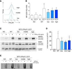
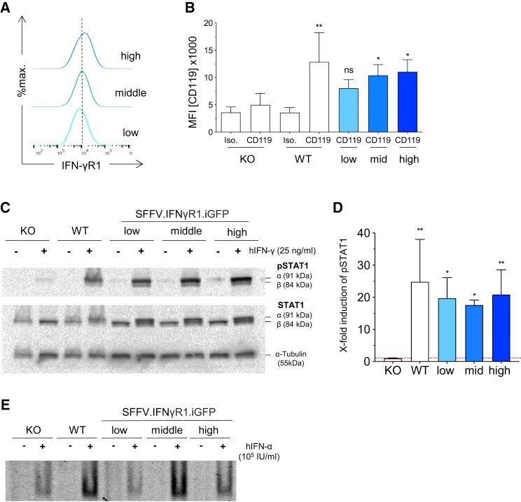
- Figure 4 Different IFN-gammaR1 Expression Levels in HeLa Cells (A) Representative flow cytometry data showing the results of FACS-purification in low, middle (mid), and high IFN-gammaR1 (CD119) expression in Lv.SFFV.IFNgammaR1.iGFP transduced KO HeLa. (B) IFN-gammaR1 (CD119) expression in different HeLa cell lines (n = 4; mean +- SD, statistical analysis was performed using one-way-ANOVA with Dunnett's multiple comparison post hoc testing. *p
- Conjugate
- Yellow dye
- Submitted by
- Invitrogen Antibodies (provider)
- Main image
- Experimental details
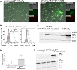
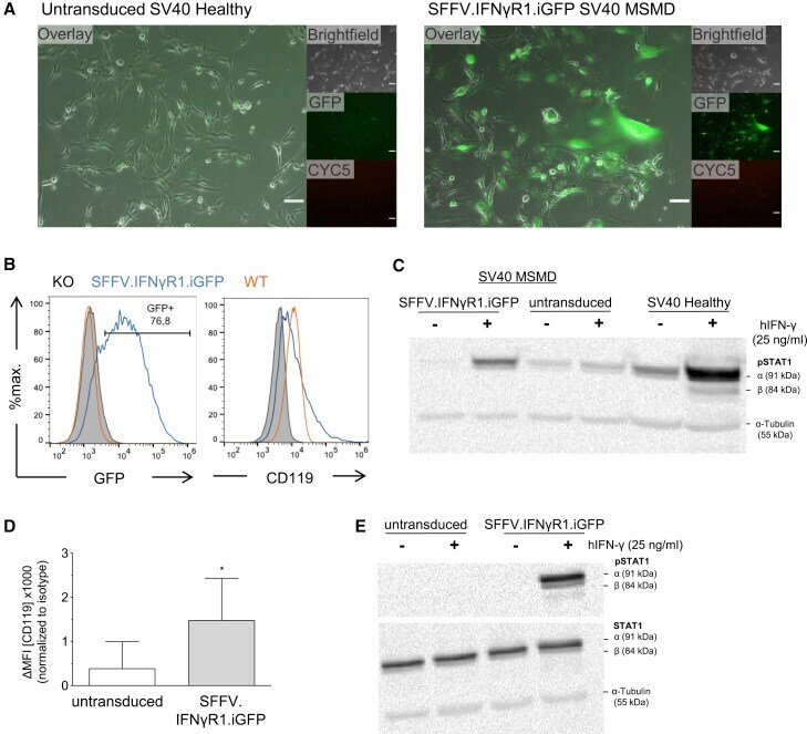
- Figure 5 Vector Analysis in SV40 Fibroblasts derived from an IFN-gammaR1 -/- MSMD Patient (A) Detection of green fluorescent protein (GFP) expression and morphology of Lv.SFFV.IFNgammaR1.iGFP-transduced SV40-immortalized fibroblasts from an IFN-gammaR1 -/- -MSMD patient by fluorescence microscopy. Untransduced SV40-immortalized fibroblasts from a healthy donor shown as control (scale bar, 100 muM). (B) Flow cytometric analysis of IFN-gammaR1 (CD119) expression in untransduced IFN-gammaR1 -/- -MSMD SV40-immortalized fibroblasts (gray filled), healthy SV40 fibroblasts (orange), and Lv.SFFV.IFNgammaR1.iGFP-transduced IFN-gammaR1 -/- -MSMD SV40 fibroblasts (blue). (C) Western blot analysis of pSTAT1 upon stimulation with 25 ng/mL hIFN-gamma in untransduced SV40-immortalized fibroblasts from a healthy control, untransduced, and Lv.SFFV.IFNgammaR1.iGFP-transduced SV40-immortalized fibroblasts from an IFN-gammaR1 -/- -MSMD patient with alpha-tubulin as loading control. (D) DeltaMean fluorescence intensity (MFI; corrected to isotype) of IFN-gammaR1 (CD119) expression in corrected and untransduced patient fibroblasts (n = 8 for untransduced cells, n = 4 for transduced cells, mean +- SD; statistical analysis was performed using unpaired t test, *p
- Conjugate
- Yellow dye
- Submitted by
- Invitrogen Antibodies (provider)
- Main image
- Experimental details
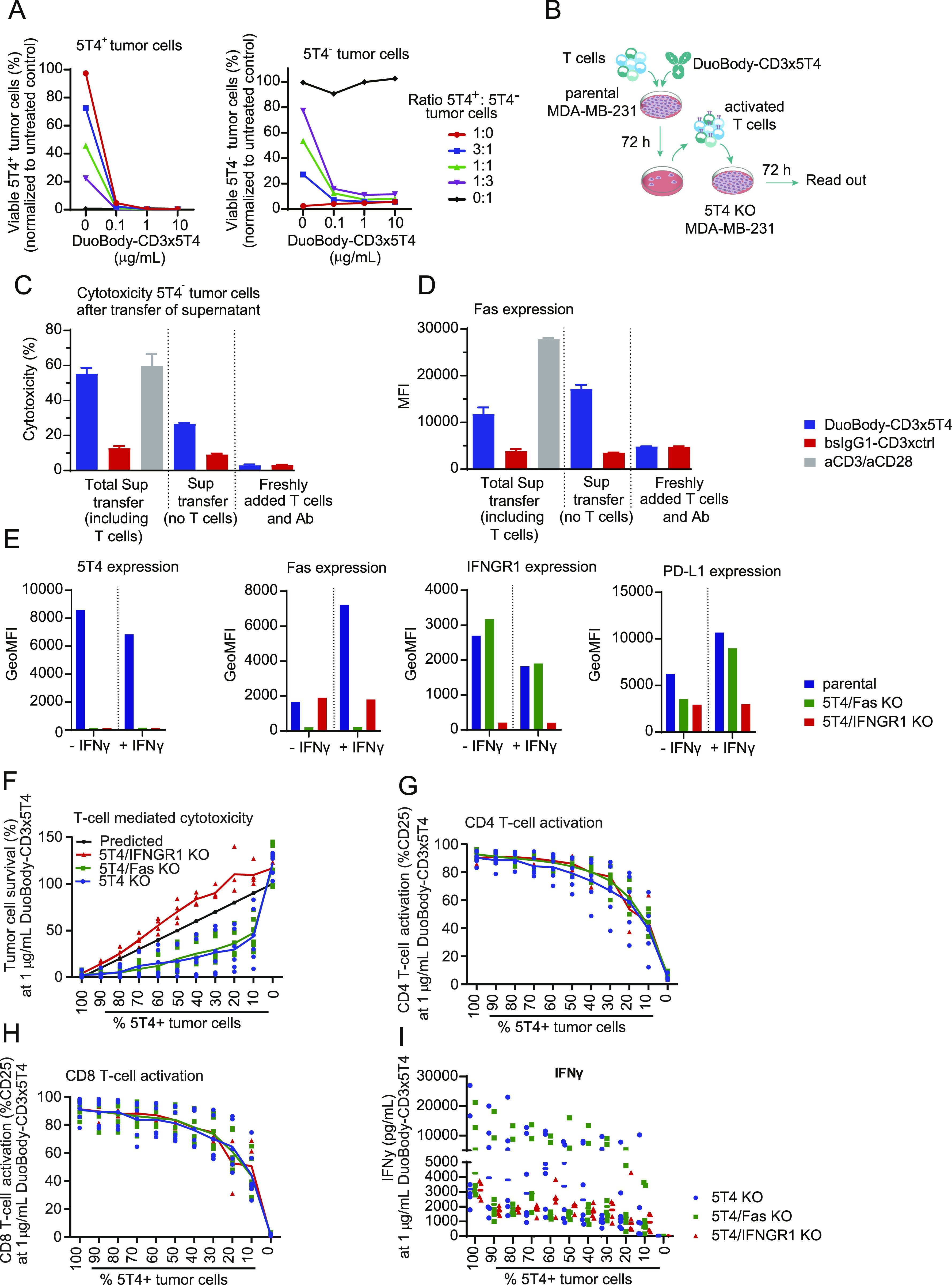
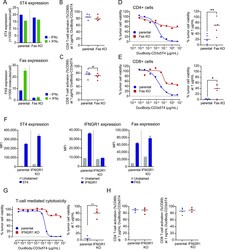
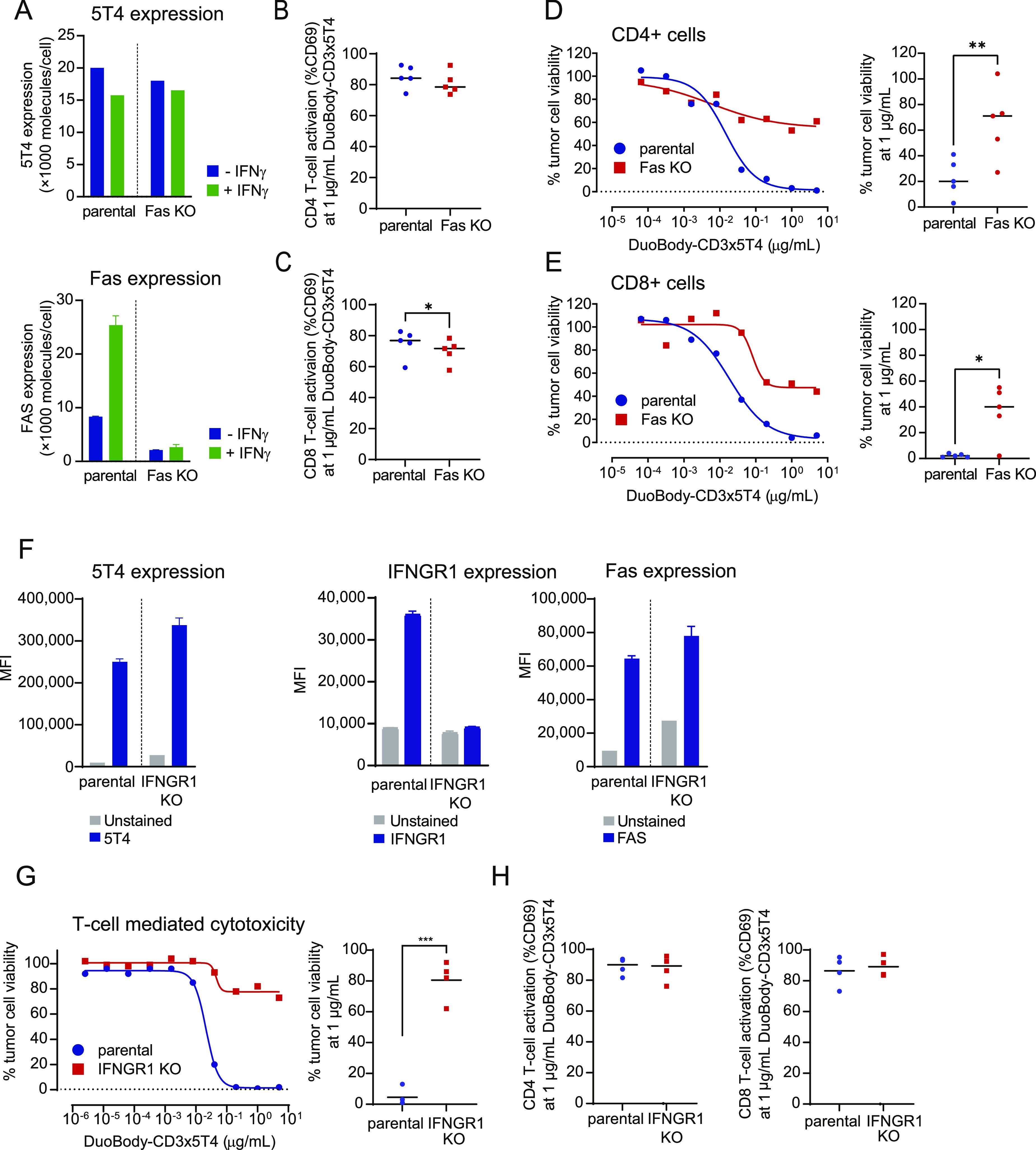
- Figure 3. Fas and IFNGR1 expression contribute to the induction of T cell-mediated cytotoxicity by DuoBody-CD3x5T4. (A) 5T4 and Fas expression on MDA-MB-231 parental and Fas KO cells was measured by quantitative flow cytometry with or without prior overnight incubation with IFNgamma (100 ng/ml). (B, C, D, E) MDA-MB-231 parental and Fas KO cells were incubated with purified CD4 + (B, D) or CD8 + (C, E) T cells (E:T ratio = 4:1, n = 5 donors) and DuoBody-CD3x5T4 for 72 h. T-cell activation (B, C) and T cell-mediated cytotoxicity (D, E) were analyzed. (B, C) Activation of T cells from five donors at 1 mug/ml DuoBody-CD3x5T4 (* P
- Conjugate
- Yellow dye
- Submitted by
- Invitrogen Antibodies (provider)
- Main image
- Experimental details
- Figure 4. DuoBody-CD3x5T4 can induce bystander kill which is dependent on IFNGR1 expression. (A) Parental and 5T4 KO MDA-MB-231 tumor cells were mixed in different ratios, as indicated, and incubated with purified T cells (E:T ratio = 4:1, n = 2 donors) and DuoBody-CD3x5T4 for 72 h. T cell-mediated cytotoxicity of 5T4 + (left panel) and 5T4 - (right panel) tumor cells was determined by flow cytometry, showing a representative donor of three donors tested. (B, C, D) Parental (5T4 + ) MDA-MB-231 tumor cells were cocultured with purified T cells (E:T = 4:1, n = 2 donors) and incubated with 10 mug/ml DuoBody-CD3x5T4 or bsIgG1-CD3xctrl for 72 h. As a positive control, T cells were incubated with anti-CD3/CD28 beads (but without tumor cells) for 72 h. (B) The supernatant (either with or without T cells) was transferred to MDA-MB-231 5T4 KO cells and incubated for 72 h (B). As negative control, MDA-MB-231 5T4 KO cells were incubated with fresh T cells and indicated antibodies for 72 h. (C, D) T cell-mediated cytotoxicity (C) and Fas expression (D) of MDA-MB-231 5T4 KO cells were determined by flow cytometry. A representative donor of two donors tested is shown. (E) 5T4, Fas, IFNGR1, and PD-L1 expression on MDA-MB-231 parental, 5T4/Fas KO, and 5T4/IFNGR1 KO cells was measured by flow cytometry with or without prior overnight incubation with IFNgamma (100 ng/ml). (F, G, H, I) Parental and 5T4/Fas KO or 5T4/IFNGR1 MDA-MB-231 tumor cells were mixed in different ratios, as indicated, and
- Conjugate
- Yellow dye