 Explore
 Validate
 Learn
Learn Western blot
Antibody data
- Antibody Data
- Antigen structure
- References [1]
- Comments [0]
- Validations
- Western blot [4]
- Immunocytochemistry [2]
- Immunohistochemistry [1]
Submit
Validation data
Reference
Comment
Report error
- Product number
- PA5-27841 - Provider product page

- Provider
- Invitrogen Antibodies
- Product name
- IFNGR1 Polyclonal Antibody
- Antibody type
- Polyclonal
- Antigen
- Recombinant protein fragment
- Description
- Recommended positive controls: HeLa, THP-1, HepG2, HepG2 membrane extract. Predicted reactivity: Rhesus Monkey (95%). Store product as a concentrated solution. Centrifuge briefly prior to opening the vial.
- Reactivity
- Human
- Host
- Rabbit
- Isotype
- IgG
- Vial size
- 100 µL
- Concentration
- 0.5 mg/mL
- Storage
- Store at 4°C short term. For long term storage, store at -20°C, avoiding freeze/thaw cycles.
Submitted references Loss of ELF5-FBXW7 stabilizes IFNGR1 to promote the growth and metastasis of triple-negative breast cancer through interferon-γ signalling.
Singh S, Kumar S, Srivastava RK, Nandi A, Thacker G, Murali H, Kim S, Baldeon M, Tobias J, Blanco MA, Saffie R, Zaidi MR, Sinha S, Busino L, Fuchs SY, Chakrabarti R
Nature cell biology 2020 May;22(5):591-602
Nature cell biology 2020 May;22(5):591-602
No comments: Submit comment
Supportive validation
- Submitted by
- Invitrogen Antibodies (provider)
- Main image
- Experimental details
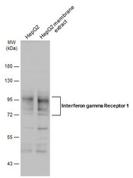
- Western Blot using IFNGR1 Polyclonal Antibody (Product # PA5-27841). HepG2 whole cell and membrane extracts (30 µg) were separated by 10% SDS-PAGE, and the membrane was blotted with IFNGR1 Polyclonal Antibody (Product # PA5-27841) diluted at 1:1,000. The HRP-conjugated anti-rabbit IgG antibody was used to detect the primary antibody.

- Submitted by
- Invitrogen Antibodies (provider)
- Main image
- Experimental details
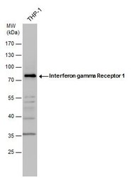
- IFNGR1 Polyclonal Antibody detects Interferon gamma Receptor 1 protein by western blot analysis. Whole cell extracts (30 µg) was separated by 10% SDS-PAGE, and the membrane was blotted with IFNGR1 Polyclonal Antibody (Product # PA5-27841) diluted by 1:1,000. The HRP-conjugated anti-rabbit IgG antibody was used to detect the primary antibody.

- Submitted by
- Invitrogen Antibodies (provider)
- Main image
- Experimental details
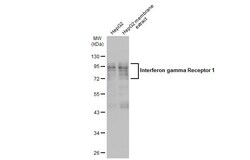
- Western Blot analysis of IFNGR1 was performed by separating 30 µg of HepG2 whole cell and membrane extracts by 7.5% SDS-PAGE. Proteins were transferred to a membrane and probed with a IFNGR1 Polyclonal Antibody (Product # PA5-27841) at a dilution of 1:1000 and a HRP-conjugated anti-rabbit IgG secondary antibody.

- Submitted by
- Invitrogen Antibodies (provider)
- Main image

- Experimental details
- Western blot analysis was performed on Membrane enriched cell extracts (30 µg lysate) of THP-1 (Lane 1), THP-1 treated with PMA (100nmol/L for 3 days followed by PMA-free medium for 1 day) (Lane 2), HeLa (Lane 3), HepG2 (Lane 4), MCF7 (Lane 5) and Caco2 (Lane 6). The blot was probed with Anti-IFNGR1 Polyclonal Antibody (Product # PA5-27841, 1:1000 dilution) and detected by chemiluminescence using Goat anti-Rabbit IgG (H+L) Superclonal™ Secondary Antibody, HRP conjugate (Product # A27036, 0.25 µg/ml, 1:4000 dilution). A 75kDa band corresponding to IFNGR1 was observed across all the cell lines tested and was observed to be induced upon treatment of THP-1 cells with PMA. An additional band was also observed at ~52kDa.

Supportive validation
- Submitted by
- Invitrogen Antibodies (provider)
- Main image

- Experimental details
- IFNGR1 Polyclonal Antibody detects Interferon gamma Receptor 1 protein at cell membrane by immunofluorescent analysis. Sample: HepG2 cells were fixed in 4% paraformaldehyde at RT for 15 min. Green: Interferon gamma Receptor 1 stained by IFNGR1 Polyclonal Antibody (Product # PA5-27841) diluted at 1:500. Blue: Fluoroshield with DAPI .

- Submitted by
- Invitrogen Antibodies (provider)
- Main image

- Experimental details
- Immunofluorescence analysis of IFNGR1 was performed using 70% confluent log phase THP-1 cells. The cells were fixed with 4% paraformaldehyde for 10 minutes, permeabilized with 0.1% Triton™ X-100 for 15 minutes, and blocked with 1% BSA for 1 hour at room temperature. The cells were labeled with IFNGR1 Polyclonal Antibody (Product # PA5-27841) at 1:100 dilution in 0.1% BSA, incubated at 4 degree Celsius overnight and then labeled with Goat anti-Rabbit IgG (H+L) Superclonal™ Secondary Antibody, Alexa Fluor® 488 conjugate (Product # A27034) at a dilution of 1:2000 for 45 minutes at room temperature (Panel a: green). Nuclei (Panel b: blue) were stained with ProLong™ Diamond Antifade Mountant with DAPI (Product # P36962). F-actin (Panel c: red) was stained with Rhodamine Phalloidin (Product # R415). Panel d represents the merged image showing membrane localization. Panel e represents control cells with no primary antibody to assess background. The images were captured at 60X magnification.

Supportive validation
- Submitted by
- Invitrogen Antibodies (provider)
- Main image
- Experimental details
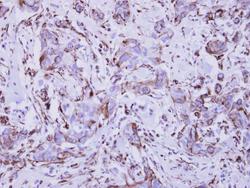
- Immunohistochemical analysis of paraffin-embedded human breast cancer, using Interferon gamma Receptor 1 (Product # PA5-27841) antibody at 1:250 dilution. Antigen Retrieval: EDTA based buffer, pH 8.0, 15 min.
