 Explore
 Validate
 Learn
Learn Western blot
Antibody data
- Antibody Data
- Antigen structure
- References [3]
- Comments [0]
- Validations
- Western blot [2]
- ELISA [1]
- Immunocytochemistry [1]
- Immunohistochemistry [1]
Submit
Validation data
Reference
Comment
Report error
- Product number
- H00007520-M02 - Provider product page

- Provider
- Novus Biologicals
- Proper citation
- Novus Cat#H00007520-M02, RRID:AB_549185
- Product name
- Mouse Monoclonal Ku80/XRCC5 Antibody
- Antibody type
- Monoclonal
- Description
- IgG purified. XRCC5 - X-ray repair complementing defective repair in Chinese hamster cells 5 (double-strand-break rejoining
- Reactivity
- Human
- Host
- Mouse
- Isotype
- IgG
- Vial size
- 0.1 mg
- Storage
- Aliquot and store at -20C or -80C. Avoid freeze-thaw cycles.
Submitted references Influence of XRCC4 expression in esophageal cancer cells on the response to radiotherapy.
Expression of Ku70 predicts results of radiotherapy in prostate cancer.
Serum anti-Ku86 is a potential biomarker for early detection of hepatitis C virus-related hepatocellular carcinoma.
Hori M, Someya M, Matsumoto Y, Nakata K, Kitagawa M, Hasegawa T, Tsuchiya T, Fukushima Y, Gocho T, Sato Y, Ohnuma H, Kato J, Sugita S, Hasegawa T, Sakata KI
Medical molecular morphology 2017 Mar;50(1):25-33
Medical molecular morphology 2017 Mar;50(1):25-33
Expression of Ku70 predicts results of radiotherapy in prostate cancer.
Hasegawa T, Someya M, Hori M, Matsumoto Y, Nakata K, Nojima M, Kitagawa M, Tsuchiya T, Masumori N, Hasegawa T, Sakata KI
Strahlentherapie und Onkologie : Organ der Deutschen Rontgengesellschaft ... [et al] 2017 Jan;193(1):29-37
Strahlentherapie und Onkologie : Organ der Deutschen Rontgengesellschaft ... [et al] 2017 Jan;193(1):29-37
Serum anti-Ku86 is a potential biomarker for early detection of hepatitis C virus-related hepatocellular carcinoma.
Nomura F, Sogawa K, Noda K, Seimiya M, Matsushita K, Miura T, Tomonaga T, Yoshitomi H, Imazeki F, Takizawa H, Mogushi K, Miyazaki M, Yokosuka O
Biochemical and biophysical research communications 2012 May 18;421(4):837-43
Biochemical and biophysical research communications 2012 May 18;421(4):837-43
No comments: Submit comment
Supportive validation
- Submitted by
- Novus Biologicals (provider)
- Main image
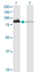
- Experimental details
- Western Blot: Ku80/XRCC5 Antibody (3D8) [H00007520-M02] - Analysis of XRCC5 expression in transfected 293T cell line by XRCC5 monoclonal antibody (M02), clone 3D8.Lane 1: XRCC5 transfected lysate(82.7 KDa).Lane 2: Non-transfected lysate.
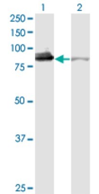
- Submitted by
- Novus Biologicals (provider)
- Main image
- Experimental details
- Western Blot: Ku80/XRCC5 Antibody (3D8) [H00007520-M02] - XRCC5 monoclonal antibody (M02), clone 3D8. Analysis of XRCC5 expression in A-431.
Supportive validation
- Submitted by
- Novus Biologicals (provider)
- Main image
- Experimental details
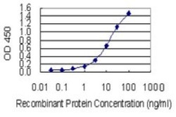
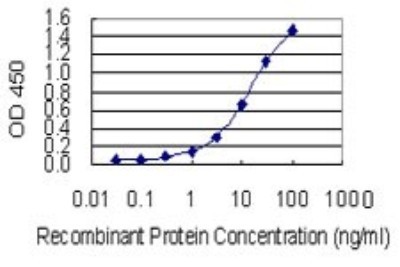
- Sandwich ELISA: Ku80/XRCC5 Antibody (3D8) [H00007520-M02] - Detection limit for recombinant GST tagged XRCC5 is 0.3 ng/ml as a capture antibody.
Supportive validation
- Submitted by
- Novus Biologicals (provider)
- Main image

- Experimental details
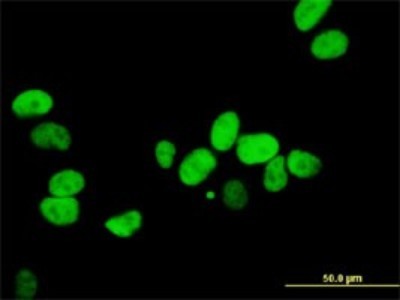
- Immunocytochemistry/Immunofluorescence: Ku80/XRCC5 Antibody (3D8) [H00007520-M02] - Analysis of monoclonal antibody to XRCC5 on HeLa cell . Antibody concentration 10 ug/ml.
Supportive validation
- Submitted by
- Novus Biologicals (provider)
- Main image
- Experimental details
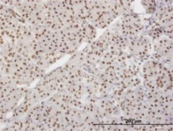
- Immunohistochemistry-Paraffin: Ku80/XRCC5 Antibody (3D8) [H00007520-M02] - Analysis of monoclonal antibody to XRCC5 on formalin-fixed paraffin-embedded human pancreas. Antibody concentration 3 ug/ml.
