 Explore
 Validate
 Learn
Learn Immunocytochemistry
 Immunoprecipitation
Antibody data
- Antibody Data
- Antigen structure
- References [0]
- Comments [0]
- Validations
- Immunocytochemistry [4]
Submit
Validation data
Reference
Comment
Report error
- Product number
- MA1-10390 - Provider product page

- Provider
- Invitrogen Antibodies
- Product name
- Ku80 Monoclonal Antibody (MEM-54)
- Antibody type
- Monoclonal
- Antigen
- Other
- Description
- This antibody reacts with Ku80, a 80 kDa subunit of Ku autoantigen (heterodimer of 72 and 87 kDa intracellular polypeptides). Ku autoantigen is involved in DNA repair and in V(D)J recombination through its ability to bind DNA double-strand breaks. Immunocytochemistry: PFA-fixation possible.
- Reactivity
- Human
- Host
- Mouse
- Isotype
- IgG
- Antibody clone number
- MEM-54
- Vial size
- 100 µg
- Concentration
- 1.0 mg/mL
- Storage
- 4° C, do not freeze
No comments: Submit comment
Supportive validation
- Submitted by
- Invitrogen Antibodies (provider)
- Main image

- Experimental details
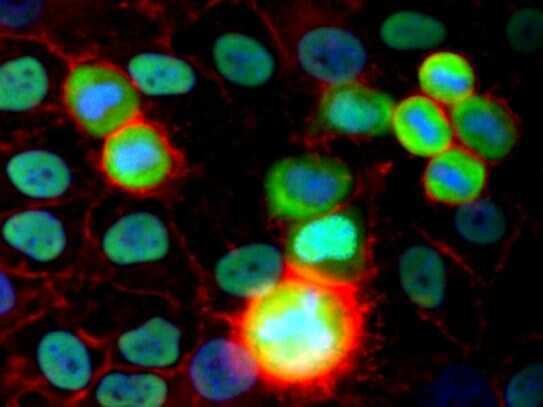
- Immunofluorescence staining of HeLa human cervix carcinoma cell line using purified anti-Ku Antigen (MEM-54) (detection by Goat anti-mouse IgG2a Alexa Fluor® 488; green). Actin (red) was stained with Phalloidin-TRITC, nuclei with DAPI (blue).
- Submitted by
- Invitrogen Antibodies (provider)
- Main image

- Experimental details
- Immunocytochemistry staining of HeLa human cervix carcinoma cell line using purified anti-Ku Antigen (MEM-54) Monoclonal antibody (Product # MA1-10390) (detection by Goat anti-mouse IgG2a Alexa Fluor® 488; green). Actin (red) was stained with Phalloidin-TRITC, nuclei with DAPI (blue).

- Submitted by
- Invitrogen Antibodies (provider)
- Main image

- Experimental details
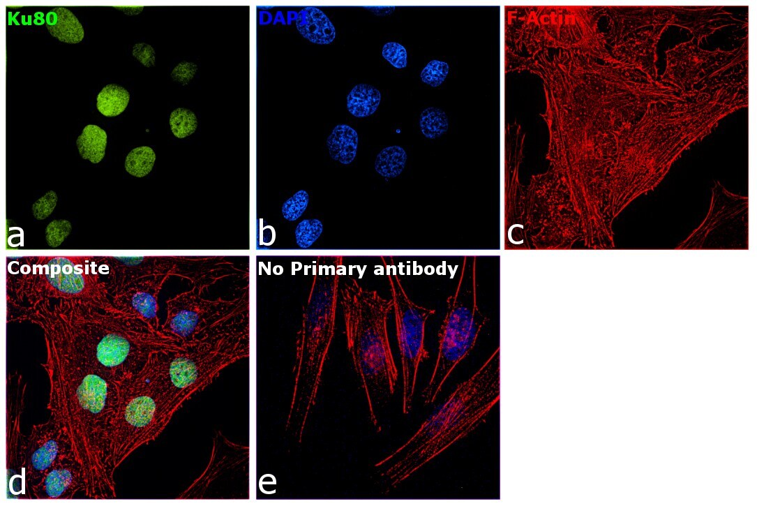
- Immunofluorescence analysis of Ku80 was performed using 70% confluent log phase HeLa cells. The cells were fixed with 4% paraformaldehyde for 10 minutes, permeabilized with 0.1% Triton™ X-100 for 15 minutes, and blocked with 2% BSA for 1 hour at room temperature. The cells were labeled with Ku80 Monoclonal Antibody (MEM-54) (Product # MA1-10390) at 1:200 dilution in 0.1% BSA, incubated at 4 degree celsius overnight and then with Donkey anti-Mouse IgG (H+L) Highly Cross-Adsorbed Secondary Antibody, Alexa Fluor Plus 488 (Product # A32766) at a dilution of 1:2000 for 45 minutes at room temperature (Panel a: green). Nuclei (Panel b: blue) were stained with SlowFade® Gold Antifade Mountant with DAPI (Product # S36938). F-actin (Panel c: red) was stained with Rhodamine Phalloidin (Product # R415, 1:300). Panel d represents the merged image showing staining in nucleus. Panel e represents control cells with no primary antibody to assess background. The images were captured at 60X magnification.
- Submitted by
- Invitrogen Antibodies (provider)
- Main image

- Experimental details
- Knockdown of Ku80 was achieved by transfecting HeLa cells with Ku80 specific siRNA (Silencer® select Product # s14952, s14953). Immunofluorescence analysis was performed on untransfected HeLa cells (panel a,d), transfected with non-specific scrambled siRNA (panels b,e) and transfected with Ku80 specific siRNA (panel c,f). Cells were fixed, permeabilized, and labelled with Ku80 Monoclonal Antibody (MEM-54) (Product # MA1-10390, 1:200 dilution) followed by Donkey anti-Mouse IgG (H+L) Highly Cross-Adsorbed Secondary Antibody, Alexa Fluor Plus 488 (Product # A32766, (1:2000 dilution). Nuclei (blue) were stained with SlowFade® Gold Antifade Mountant with DAPI (Product # S36938), and Rhodamine Phalloidin (Product # R415, 1:300) was used for cytoskeletal F-actin (Red) staining. Reduction of specific signal was observed upon siRNA mediated knockdown (panel c,f) confirming specificity of the antibody to Ku80 (Green). The Images were captured at 60X magnification.
