Explore
 Validate
 Learn
Learn Western blot
 ELISA
ELISA Immunocytochemistry
Antibody data
- Antibody Data
- Antigen structure
- References [1]
- Comments [0]
- Validations
- Immunocytochemistry [2]
Submit
Validation data
Reference
Comment
Report error
- Product number
- PA5-89161 - Provider product page

- Provider
- Invitrogen Antibodies
- Product name
- LRP6 Polyclonal Antibody
- Antibody type
- Polyclonal
- Antigen
- Recombinant full-length protein
- Description
- Immunogen sequence: APLLLYANRR DLRLVDATNG KENATIVVGG LEDAAAVDFV FSHGLIYWSD VSEEAIKRTE FNKTESVQNV VVSGLLSPDG LACDWLGEKL YWTDSETNRI EVSNLDGSLR KVLFWQELDQ PRAIALDPSS G; Positive Samples: Mouse liver; Cellular Location: Endoplasmic reticulum, Membrane, Single-pass type I membrane protein
- Reactivity
- Human, Mouse
- Host
- Rabbit
- Isotype
- IgG
- Vial size
- 100 μL
- Concentration
- 2.14 mg/mL
- Storage
- -20°C, Avoid Freeze/Thaw Cycles
Submitted references HLY78 Attenuates Neuronal Apoptosis via the LRP6/GSK3β/β-Catenin Signaling Pathway After Subarachnoid Hemorrhage in Rats.
Luo X, Li L, Xu W, Cheng Y, Xie Z
Neuroscience bulletin 2020 Oct;36(10):1171-1181
Neuroscience bulletin 2020 Oct;36(10):1171-1181
No comments: Submit comment
Supportive validation
- Submitted by
- Invitrogen Antibodies (provider)
- Main image

- Experimental details
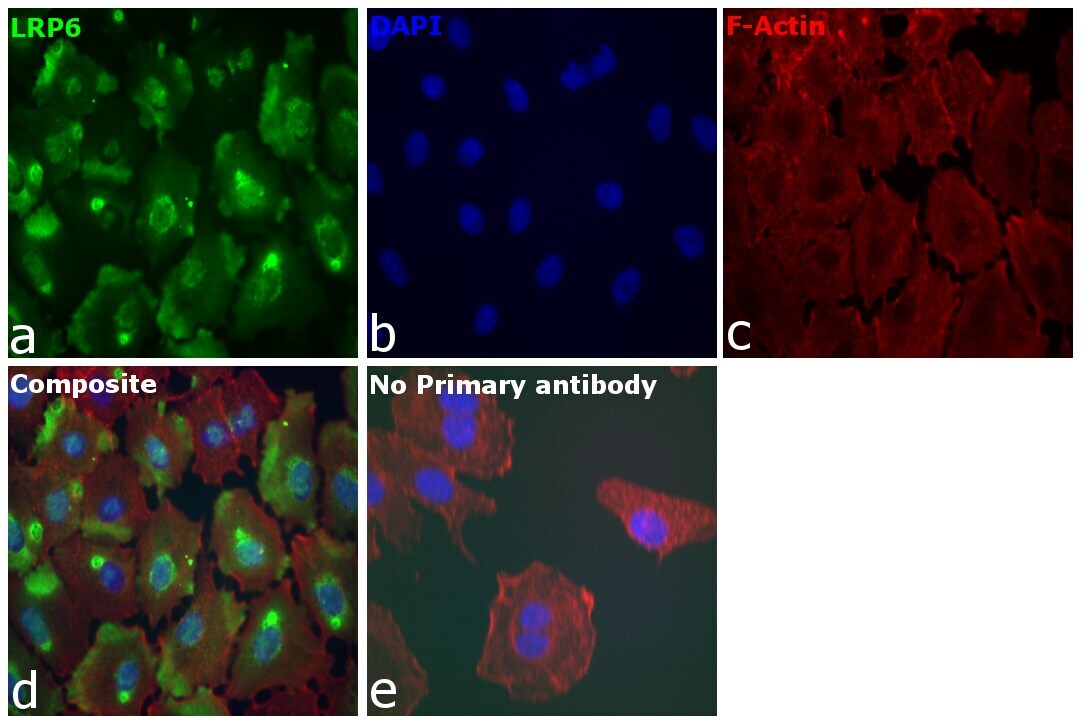
- Immunofluorescence analysis of LRP6 was performed using 70% confluent log phase A549 cells. The cells were fixed with 4% paraformaldehyde for 10 minutes, permeabilized with 0.01% Triton™ X-100 for 15 minutes, and blocked with 2% BSA for 10 minutes at room temperature. The cells were labeled with LRP6 Polyclonal Antibody (Product # PA5-89161) at 1:100 dilution in 0.1% BSA, incubated at 4 degrees celsius overnight, and then labeled with Donkey anti-Rabbit IgG (H+L) Highly Cross-Adsorbed Secondary Antibody, Alexa Fluor Plus 488 (Product # A32790), (1:2000 dilution), for 45 minutes at room temperature (Panel a: Green). Nuclei (Panel b: Blue) were stained with Hoechst 33342 (Product # H1399). F-actin (Panel c: Red) was stained with Alexa Fluor™ Plus 647 Phalloidin (Product # A30107, 1:2000 dilution). Panel d represents the merged image showing endoplasmic reticulum localization. Panel e represents control cells with no primary antibody to assess the background. The images were captured at 40X magnification in CellInsight CX7 LZR High-Content Screening (HCS) Platform (Product # CX7A1110LZR).

- Submitted by
- Invitrogen Antibodies (provider)
- Main image

- Experimental details
- Immunofluorescence analysis of LRP6 was performed using 70% confluent log phase A549 cells. The cells were fixed with 4% paraformaldehyde for 10 minutes, permeabilized with 0.01% Triton™ X-100 for 15 minutes, and blocked with 2% BSA for 10 minutes at room temperature. The cells were labeled with LRP6 Polyclonal Antibody (Product # PA5-89161) at 1:100 dilution in 0.1% BSA, incubated at 4 degrees celsius overnight, and then labeled with Donkey anti-Rabbit IgG (H+L) Highly Cross-Adsorbed Secondary Antibody, Alexa Fluor Plus 488 (Product # A32790), (1:2000 dilution), for 45 minutes at room temperature (Panel a: Green). Nuclei (Panel b: Blue) were stained with Hoechst 33342 (Product # H1399). F-actin (Panel c: Red) was stained with Alexa Fluor™ Plus 647 Phalloidin (Product # A30107, 1:2000 dilution). Panel d represents the merged image showing endoplasmic reticulum localization. Panel e represents control cells with no primary antibody to assess the background. The images were captured at 40X magnification in CellInsight CX7 LZR High-Content Screening (HCS) Platform (Product # CX7A1110LZR).