 Explore
 Validate
 Learn
Learn Western blot
 ELISA
ELISAAntibody data
- Antibody Data
- Antigen structure
- References [5]
- Comments [0]
- Validations
- Western blot [4]
- Immunohistochemistry [1]
Submit
Validation data
Reference
Comment
Report error
- Product number
- GTX22480 - Provider product page

- Provider
- GeneTex
- Proper citation
- GeneTex Cat#GTX22480, RRID:AB_374325
- Product name
- Myosin Light chain 2 (phospho Ser19) antibody
- Antibody type
- Polyclonal
- Reactivity
- Human, Mouse
- Host
- Rabbit
Submitted references The Transcription Factor Foxg1 Promotes Optic Fissure Closure in the Mouse by Suppressing Wnt8b in the Nasal Optic Stalk.
Wnt ligands from the embryonic surface ectoderm regulate 'bimetallic strip' optic cup morphogenesis in mouse.
Balanced Rac1 and RhoA activities regulate cell shape and drive invagination morphogenesis in epithelia.
A Trio-RhoA-Shroom3 pathway is required for apical constriction and epithelial invagination.
Cdc42- and IRSp53-dependent contractile filopodia tether presumptive lens and retina to coordinate epithelial invagination.
Smith R, Huang YT, Tian T, Vojtasova D, Mesalles-Naranjo O, Pollard SM, Pratt T, Price DJ, Fotaki V
The Journal of neuroscience : the official journal of the Society for Neuroscience 2017 Aug 16;37(33):7975-7993
The Journal of neuroscience : the official journal of the Society for Neuroscience 2017 Aug 16;37(33):7975-7993
Wnt ligands from the embryonic surface ectoderm regulate 'bimetallic strip' optic cup morphogenesis in mouse.
Carpenter AC, Smith AN, Wagner H, Cohen-Tayar Y, Rao S, Wallace V, Ashery-Padan R, Lang RA
Development (Cambridge, England) 2015 Mar 1;142(5):972-82
Development (Cambridge, England) 2015 Mar 1;142(5):972-82
Balanced Rac1 and RhoA activities regulate cell shape and drive invagination morphogenesis in epithelia.
Chauhan BK, Lou M, Zheng Y, Lang RA
Proceedings of the National Academy of Sciences of the United States of America 2011 Nov 8;108(45):18289-94
Proceedings of the National Academy of Sciences of the United States of America 2011 Nov 8;108(45):18289-94
A Trio-RhoA-Shroom3 pathway is required for apical constriction and epithelial invagination.
Plageman TF Jr, Chauhan BK, Yang C, Jaudon F, Shang X, Zheng Y, Lou M, Debant A, Hildebrand JD, Lang RA
Development (Cambridge, England) 2011 Dec;138(23):5177-88
Development (Cambridge, England) 2011 Dec;138(23):5177-88
Cdc42- and IRSp53-dependent contractile filopodia tether presumptive lens and retina to coordinate epithelial invagination.
Chauhan BK, Disanza A, Choi SY, Faber SC, Lou M, Beggs HE, Scita G, Zheng Y, Lang RA
Development (Cambridge, England) 2009 Nov;136(21):3657-67
Development (Cambridge, England) 2009 Nov;136(21):3657-67
No comments: Submit comment
Supportive validation
- Submitted by
- GeneTex (provider)
- Main image
- Experimental details
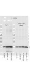
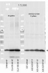
- Affinity Purified Phospho specific antibody to Monophosphorylated Regulatory Light Chain of Smooth and Non-muscle Myosin at pS19/pS20 (GTX22480) was used at a 1:5000 dilution to detect myosin light chain by Western blot. Either 13 or 20 ul of a mouse cardiac myocyte lysate was loaded on a 4-20% Criterion gel for SDS-PAGE. Samples were either mock-treated or CLA-treated, as indicated. After washing, a 1:5,000 dilution of HRP conjugated Gt-a-Rabbit IgG (GTX27090) preceded color development using Amersham's substrate system.
- Validation comment
- WB
- Submitted by
- GeneTex (provider)
- Main image
- Experimental details
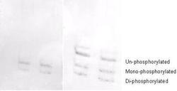
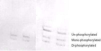
- Affinity purified phosphospecific antibody to phosphorylated regulatory light chain of smooth and non-muscle Myosin at pS19/pS20 (GTX22480) was used at a 1:1000 dilution to detect myosin light chain by Western blot on 3T3 cell lysates. A standard urea/glycerol gel without SDS was used to separate phospho forms of regulatory light chain according to mass to charge ratios. In Panel A, reactivity of GeneTex phosphospecific antibody is shown. In Panel B, reactivity of commercially available pan reactive antibody that detects both unphosphorylated and phosphorylated forms of regulatory light chain is shown. GeneTex phosphospecific antibody detects both monophosphorylated (pSer20 Mono-P-RLC) and diphosphorylated (pThr19-pSer20 Di-P-RLC) regulatory light chain. Personal communication. J. Stull. UT Southwestern Medical Center
- Validation comment
- WB
- Submitted by
- GeneTex (provider)
- Main image
- Experimental details
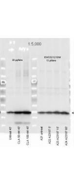
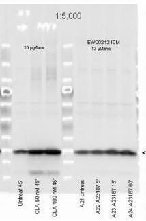
- Myosin Light chain (phospho Ser20) antibody (GTX22480) was used at a 1:5000 dilution to detect myosin light chain by Western blot. Either 13 or 20 ul of a mouse cardiac myocyte lysate was loaded on a 4-20% Criterion gel for SDS-PAGE. Samples were either mock-treated or CLA-treated, as indicated. After washing, a 1:5,000 dilution of HRP conjugated Gt-a-Rabbit IgG (GTX27090) preceded color development using Amersham's substrate system
- Submitted by
- GeneTex (provider)
- Main image
- Experimental details
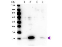
- Western blot of Myosin Light chain (phospho Ser20) antibody (GTX22480). Lane 1: Regulatory Light Chain Non-Phospho recombinant protein. Lane 2: Regulatory Light Chain Phospho recombinant protein. Lane 3: Smooth Muscle Non-Phospho recombinant protein. Lane 4: Smooth Muscle Phospho recombinant protein. Load: 50 ng per lane. Primary antibody at 1:1,000 overnight at 4¢XC. Secondary antibody: Peroxidase rabbit secondary antibody at 1:40,000 for 60 min at RT. Blocking for 30 min at RT. Predicted/Observed size: 20 kDa.
Supportive validation
- Submitted by
- GeneTex (provider)
- Main image
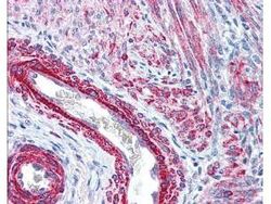
- Experimental details
- Myosin Light chain (phospho Ser20) antibody (GTX22480) was used at 2.5 ?g/ml to detect signal in a variety of tissues including multi-human, multi-brain and multi-cancer slides. This image shows strong staining of both vascular and myometrial smooth muscle cells of the uterus. Tissue was formalin-fixed and paraffin embedded. The image shows localization of the antibody as the precipitated red signal, with a hematoxylin purple nuclear counterstain.
