 Explore
 Validate
 Learn
Learn Western blot
 ELISA
ELISAAntibody data
- Antibody Data
- Antigen structure
- References [0]
- Comments [0]
- Validations
- Western blot [5]
- Immunohistochemistry [1]
Submit
Validation data
Reference
Comment
Report error
- Product number
- NB100-1687 - Provider product page

- Provider
- Novus Biologicals
- Proper citation
- Novus Cat#NB100-1687, RRID:AB_10003202
- Product name
- Rabbit Polyclonal MRCL3 Antibody
- Antibody type
- Polyclonal
- Description
- Immunogen affinity purified. Specific for the phosphorylated pS19/pS20 form of the protein, depending on the source origin of the protein. Reactivity with nonphosphorylated myosin light chain is less than 1% by ELISA. Cross reactivity is expected with myosin light chain from human and mouse.
- Reactivity
- Human, Mouse
- Host
- Rabbit
- Isotype
- IgG
- Vial size
- 0.05 mg
- Concentration
- 1 mg/ml
- Storage
- Store at -20C. Avoid freeze-thaw cycles.
No comments: Submit comment
Supportive validation
- Submitted by
- Novus Biologicals (provider)
- Main image
- Experimental details
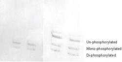
- Western Blot: MRCL3 [p Ser19, p Ser20] Antibody [NB100-1687] - Used at a 1:1000 dilution to detect myosin light chain by Western blot on 3T3 cell lysates. A standard urea/glycerol gel without SDS was used to separate phospho forms of regulatory light chain according to mass to charge ratios. In Panel A, reactivity of phosphospecific antibody is shown. In Panel B, reactivity of commercially available pan reactive antibody that detects both unphosphorylated and phosphorylated forms of regulatory light chain is shown. phosphospecific antibody detects both monophosphorylated (pSer20 Mono-P-RLC) and diphosphorylated (pThr19-pSer20 Di-P-RLC) regulatory light chain.

- Submitted by
- Novus Biologicals (provider)
- Main image
- Experimental details
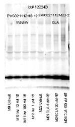
- Western Blot: MRCL3 [p Ser19, p Ser20] Antibody [NB100-1687] - Affinity purified phosphospecific antibody to monophosphorylated regulatory light chain of smooth and non-muscle Myosin at pS19 was used at a 1:1000 dilution to detect myosin light chain by Western blot. Approximately 12 ul of a mouse cardiac myocyte lysate was loaded on a 4-20% Criterion gel for SDS-PAGE. Samples were either mock treated or insulin or CLA treated according as indicated. After washing, a 1:5,000 dilution of HRP conjugated Gta- Rabbit IgG (611-103-122) preceded color development using Amersham's substrate system. Other detection methods will yield similar results.

- Submitted by
- Novus Biologicals (provider)
- Main image
- Experimental details
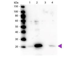
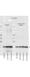
- Western Blot: MRCL3 [p Ser19, p Ser20] Antibody [NB100-1687] - Lane 1: Regulatory Light Chain Non-Phospho recombinant protein. Lane 2: Regulatory Light Chain Phospho recombinant protein. Lane 3: Smooth Muscle Non-Phospho recombinant protein. Lane 4: Smooth Muscle Phospho recombinant protein. Load: 50 ng per lane. Primary antibody: Myosin pS19/pS20 primary antibody at 1:1,000 overnight at 4C. Secondary antibody: Peroxidase rabbit secondary antibody at 1:40,000 for 60 min at RT. Blocking: incubated with blocking buffer for 30 min at RT. Predicted/Observed size: 20 kDa, 20 kDa for Regulatory Light Chain Phospho. Other band(s): None.
- Submitted by
- Novus Biologicals (provider)
- Main image
- Experimental details
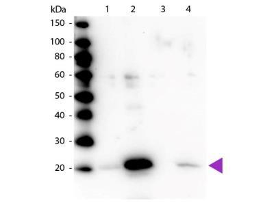
- Western Blot: MRCL3 [p Ser19, p Ser20] Antibody [NB100-1687] - Used at a 1:5000 dilution to detect myosin light chain by Western blot. Either 13 or 20 l of a mouse cardiac myocyte lysate was loaded on a 4-20% Criterion gel for SDS-PAGE. Samples were either mock-treated or CLA-treated, as indicated. After washing, a 1:5,000 dilution of HRP conjugated Gt-a-Rabbit IgG preceded color development using Amersham's substrate system.

- Submitted by
- Novus Biologicals (provider)
- Main image
- Experimental details
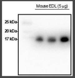
- Western Blot: MRCL3 [p Ser19, p Ser20] Antibody [NB100-1687] - This antibody has been shown to cross-react with mouse skeletal muscle regulatory light chain (Lai et al., 2016. Am J Physiol Endocrinol Metab), and is shown to positively react with mouse EDL muscle here. This image was submitted via customer Review.

Supportive validation
- Submitted by
- Novus Biologicals (provider)
- Main image
- Experimental details
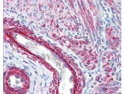
- Immunohistochemistry: MRCL3 [p Ser19, p Ser20] Antibody [NB100-1687] - Used at 2.5 ug/ml to detect signal in a variety of tissues including multi-human, multi-brain and multi-cancer slides. This image shows strong staining of both vascular and myometrial smooth muscle cells of the uterus. Tissue was formalin-fixed and paraffin embedded. The image shows localization of the antibody as the precipitated red signal, with a hematoxylin purple nuclear counterstain.
