Explore
 Validate
 Learn
Learn Western blot
 ELISA
ELISA Immunohistochemistry
Antibody data
- Antibody Data
- Antigen structure
- References [0]
- Comments [0]
- Validations
- Western blot [3]
- Immunohistochemistry [2]
Submit
Validation data
Reference
Comment
Report error
- Product number
- LS-C745286 - Provider product page

- Provider
- LSBio
- Product name
- MYL12A / MRCL3 Antibody (phospho-Ser19/20) LS-C745286
- Antibody type
- Polyclonal
- Description
- Affinity purified
- Reactivity
- Human
- Host
- Rabbit
- Isotype
- IgG
- Storage
- Store vial at -20°C or below prior to opening. Dilute 1:10 to minimize loss. Store the vial at -20°C or below after dilution. Avoid freeze-thaw cycles.
No comments: Submit comment
Enhanced validation
- Submitted by
- LSBio (provider)
- Enhanced method
- Genetic validation
- Main image

- Experimental details
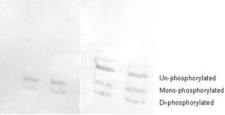
- Affinity purified phosphospecific antibody to phosphorylated regulatory light chain of smooth and non-muscle Myosin at pS19/pS20 was used at a 1:1000 dilution to detect myosin light chain by Western blot on 3T3 cell lysates. A standard urea/glycerol gel without SDS was used to separate phospho forms of regulatory light chain according to mass to charge ratios. In Panel A, reactivity of phosphospecific antibody is shown. In Panel B, reactivity of commercially available pan reactive antibody that detects both unphosphorylated and phosphorylated forms of regulatory light chain is shown. phosphospecific antibody detects both monophosphorylated (pSer20 Mono-P-RLC) and diphosphorylated (pThr19-pSer20 Di-P-RLC) regulatory light chain.
- Submitted by
- LSBio (provider)
- Enhanced method
- Genetic validation
- Main image
- Experimental details
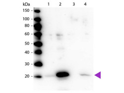
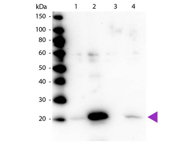
- Western blot of rabbit Anti-Myosin pS19/pS20 primary antibody. Lane 1: Regulatory Light Chain Non-Phospho recombinant protein. Lane 2: Regulatory Light Chain Phospho recombinant protein. Lane 3: Smooth Muscle Non-Phospho recombinant protein. Lane 4: Smooth Muscle Phospho recombinant protein. Load: 50 ng per lane. Primary antibody: Myosin pS19/pS20 primary antibody at 1:1,000 overnight at 4°C. Secondary antibody: Peroxidase rabbit secondary antibody at 1:40,000 for 60 min at RT. Blocking: MB-070 for 30 min at RT. Predicted/Observed size: 20 kDa, 20 kDa for Regulatory Light Chain Phospho. Other band(s): None.
- Submitted by
- LSBio (provider)
- Enhanced method
- Genetic validation
- Main image

- Experimental details
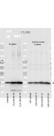
- Affinity Purified Phospho specific antibody to Monophosphorylated Regulatory Light Chain of Smooth and Non-muscle Myosin at pS19/pS20 was used at a 1:5000 dilution to detect myosin light chain by Western blot. Either 13 or 20 µl of a mouse cardiac myocyte lysate was loaded on a 4-20% Criterion gel for SDS-PAGE. Samples were either mock-treated or CLA-treated, as indicated. After washing, a 1:5,000 dilution of HRP conjugated Gt-a-Rabbit IgG preceded color development using Amersham's substrate system. Other detection methods will yield similar results. Data courtesy of the Alliance for Cellular Signaling (http://www.signaling-gateway.org).
Supportive validation
- Submitted by
- LSBio (provider)
- Main image
- Experimental details
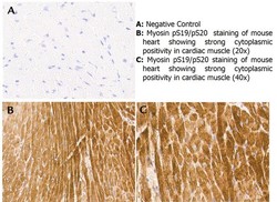
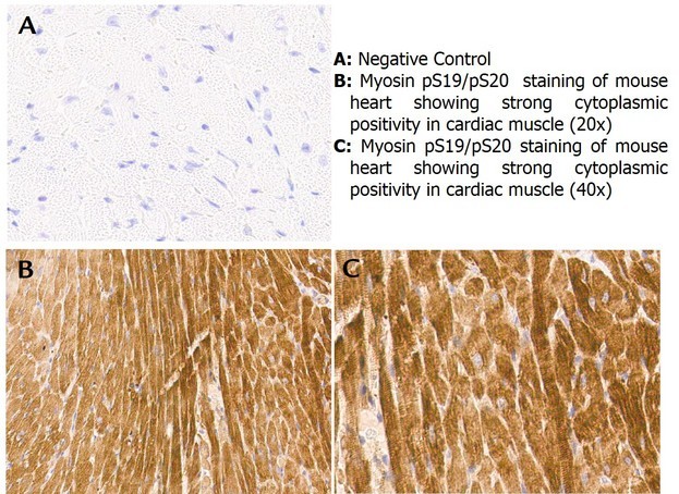
- Immunohistochemistry with anti-myosin pS19/pS20 antibody showing strong cytoplasmic staining of myocytes in mouse heart muscle 20x and 40x (B & C). Staining was performed on Leica Bond system using the standard protocol. Formalin fixed/paraffin embedded tissue sections were subjected to antigen retrieval and then incubated with rabbit anti-myosin pS19/pS20 antibody at 1:100 dilution for 60 minutes. Biotinylated Anti-rabbit secondary antibody was used to detect primary antibody. The reaction was developed using streptavidin-HRP conjugated compact polymer system and visualized with chromogen substrate, 3’3-diamino-benzidine substrate (DAB). The sections were then counterstained with hematoxylin to detect cell nuclei.
- Submitted by
- LSBio (provider)
- Main image
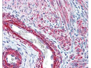
- Experimental details
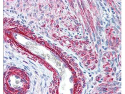
- Affinity purified anti-Monophosphorylated RLC Smooth and Non-Muscle Myosin pS19/20 antibody was used at 2.5 µg/ml to detect signal in a variety of tissues including multi-human, multi-brain and multi-cancer slides. This image shows strong staining of both vascular and myometrial smooth muscle cells of the uterus. Tissue was formalin-fixed and paraffin embedded. The image shows localization of the antibody as the precipitated red signal, with a hematoxylin purple nuclear counterstain.