Explore
 Validate
 Learn
Learn Western blot
 ELISA
ELISAAntibody data
- Antibody Data
- Antigen structure
- References [0]
- Comments [0]
- Validations
- Western blot [3]
- Immunohistochemistry [2]
Submit
Validation data
Reference
Comment
Report error
- Product number
- 600-401-416 - Provider product page

- Provider
- Invitrogen Antibodies
- Product name
- Phospho-Myosin (Ser19, Ser20) Polyclonal Antibody
- Antibody type
- Polyclonal
- Antigen
- Synthetic peptide
- Reactivity
- Human
- Host
- Rabbit
- Isotype
- IgG
- Vial size
- 100 µg
- Concentration
- 1.1 mg/mL
- Storage
- -20° C, Avoid Freeze/Thaw Cycles
No comments: Submit comment
Supportive validation
- Submitted by
- Invitrogen Antibodies (provider)
- Main image
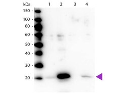
- Experimental details
- Western blot of Rabbit Anti-Myosin pS19/pS20 primary antibody. Lane 1: Regulatory Light Chain Non-Phospho recombinant protein. Lane 2: Regulatory Light Chain Phospho recombinant protein. Lane 3: Smooth Muscle Non-Phospho recombinant protein. Lane 4: Smooth Muscle Phospho recombinant protein. Load: 50 ng per lane. Primary antibody: Myosin pS19/pS20 primary antibody at 1:1,000 overnight at 4°C. Secondary antibody: Peroxidase rabbit secondary antibody at 1:40,000 for 60 min at RT. Blocking: MB-070 for 30 min at RT. Predicted/Observed size: 20 kDa, 20 kDa for Regulatory Light Chain Phospho. Other band(s): None.
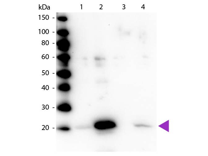
- Submitted by
- Invitrogen Antibodies (provider)
- Main image

- Experimental details
- Affinity Purified Phospho specific antibody to Monophosphorylated Regulatory Light Chain of Smooth and Non-muscle Myosin at pS19/pS20 was used at a 1:5000 dilution to detect myosin light chain by Western blot. Either 13µL or 20 µg of a mouse cardiac myocyte lysate was loaded on a 4-20% Criterion gel for SDS-PAGE. Samples were either mock-treated or CLA-treated, as indicated. After washing, a 1:5,000 dilution of HRP conjugated Gt-a-Rabbit IgG (611-103-122) preceded color development using Amershams substrate system. Other detection methods will yield similar results. Data courtesy of the Alliance for Cellular Signaling (http://www.signaling-gateway.org).
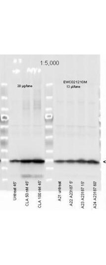
- Submitted by
- Invitrogen Antibodies (provider)
- Main image

- Experimental details
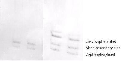
- Affinity purified phosphospecific antibody to phosphorylated regulatory light chain of smooth and non-muscle Myosin at pS19/pS20 was used at a 1:1000 dilution to detect myosin light chain by Western blot on 3T3 cell lysates. A standard urea/glycerol gel without SDS was used to separate phospho forms of regulatory light chain according to mass to charge ratios. In Panel A on the left, reactivity of Rocklands phosphospecific antibody is shown. In Panel B on the right, reactivity of commercially available pan reactive antibody that detects both un-phosphorylated and phosphorylated forms of regulatory light chain is shown. Rocklands phosphospecific antibody detects both mono-phosphorylated (pSer20 Mono-P-RLC) and di-phosphorylated (pThr19-pSer20 Di-P-RLC) regulatory light chain. Personal communication. J. Stull. UT Southwestern Medical Center.
Supportive validation
- Submitted by
- Invitrogen Antibodies (provider)
- Main image
- Experimental details
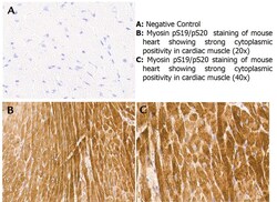
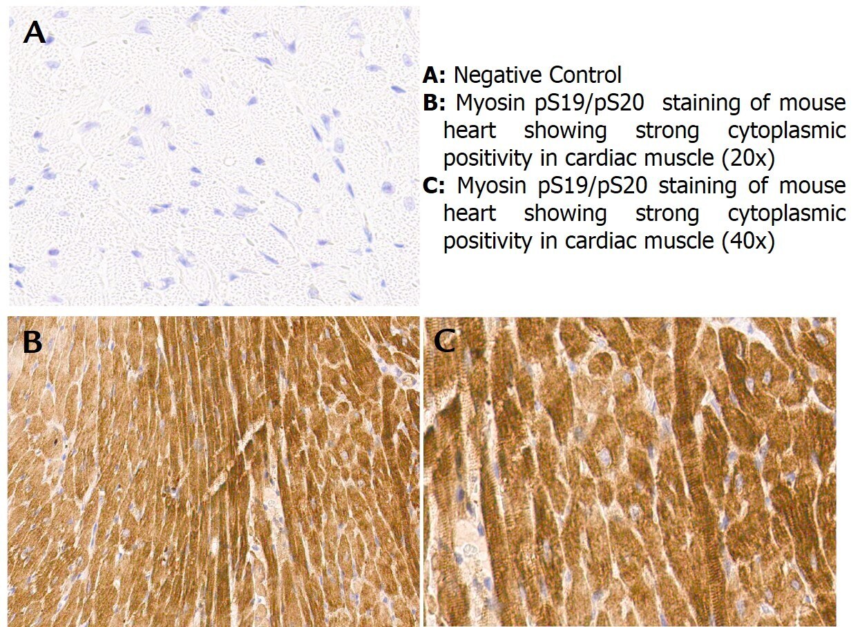
- Immunohistochemistry with anti-myosin pS19/pS20 antibody showing strong cytoplasmic staining of myocytes in mouse heart muscle 20x and 40x (B & C). Staining was performed on Leica Bond system using the standard protocol. Formalin fixed/paraffin embedded tissue sections were subjected to antigen retrieval and then incubated with rabbit anti-myosin pS19/pS20 antibody at 1:100 dilution for 60 minutes. Biotinylated Anti-rabbit secondary antibody was used to detect primary antibody. The reaction was developed using streptavidin-HRP conjugated compact polymer system and visualized with chromogen substrate, 33-diamino-benzidine substrate (DAB). The sections were then counterstained with hematoxylin to detect cell nuclei.
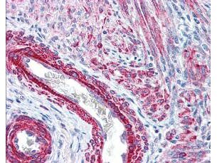
- Submitted by
- Invitrogen Antibodies (provider)
- Main image

- Experimental details
- Rocklands affinity purified anti-Monophosphorylated RLC Smooth and Non-Muscle Myosin pS19/20 antibody was used at 2.5 µg/ml to detect signal in a variety of tissues including multi-human, multi-brain and multi-cancer slides. This image shows strong staining of both vascular and myometrial smooth muscle cells of the uterus. Tissue was formalin-fixed and paraffin embedded. The image shows localization of the antibody as the precipitated red signal, with a hematoxylin purple nuclear counterstain. Personal Communication, Tina Roush, LifeSpanBiosciences, Seattle, WA.