Explore
 Validate
 Learn
Learn Western blot
 ELISA
ELISAAntibody data
- Antibody Data
- Antigen structure
- References [4]
- Comments [0]
- Validations
- Western blot [2]
- Immunohistochemistry [1]
Submit
Validation data
Reference
Comment
Report error
- Product number
- R1535P - Provider product page

- Provider
- Acris Antibodies GmbH
- Proper citation
- Acris Antibodies GmbH Cat#R1535P, RRID:AB_1005524
- Product name
- anti MYL12A pSer19/20
- Antibody type
- Polyclonal
- Antigen
- Human Myosin Light Chain phospho peptide corresponding to a region of the Human smooth/non-muscle form of Myosin regulatory light chain conjugated to Keyhole Limpet Hemocyanin (KLH).
- Reactivity
- Human
- Host
- Rabbit
- Vial size
- 0.1 mg
- Concentration
- 1.0 mg/ml (by UV absorbance at 280 nm)
Submitted references Adenosine A1 receptor-dependent and independent pathways in modulating renal vascular responses to angiotensin II.
Noradrenaline enhances angiotensin II responses via p38 MAPK activation after hypoxia/re-oxygenation in renal interlobar arteries.
Adenosine enhances long term the contractile response to angiotensin II in afferent arterioles.
Adenosine restores angiotensin II-induced contractions by receptor-independent enhancement of calcium sensitivity in renal arterioles.
Gao X, Peleli M, Zollbrecht C, Patzak A, Persson AE, Carlström M
Acta physiologica (Oxford, England) 2015 Jan;213(1):268-76
Acta physiologica (Oxford, England) 2015 Jan;213(1):268-76
Noradrenaline enhances angiotensin II responses via p38 MAPK activation after hypoxia/re-oxygenation in renal interlobar arteries.
Kaufmann J, Martinka P, Moede O, Sendeski M, Steege A, Fähling M, Hultström M, Gaestel M, Moraes-Silva IC, Nikitina T, Liu ZZ, Zavaritskaya O, Patzak A
Acta physiologica (Oxford, England) 2015 Jan 16;
Acta physiologica (Oxford, England) 2015 Jan 16;
Adenosine enhances long term the contractile response to angiotensin II in afferent arterioles.
Patzak A, Lai EY, Fähling M, Sendeski M, Martinka P, Persson PB, Persson AE
American journal of physiology. Regulatory, integrative and comparative physiology 2007 Dec;293(6):R2232-42
American journal of physiology. Regulatory, integrative and comparative physiology 2007 Dec;293(6):R2232-42
Adenosine restores angiotensin II-induced contractions by receptor-independent enhancement of calcium sensitivity in renal arterioles.
Lai EY, Martinka P, Fähling M, Mrowka R, Steege A, Gericke A, Sendeski M, Persson PB, Persson AE, Patzak A
Circulation research 2006 Nov 10;99(10):1117-24
Circulation research 2006 Nov 10;99(10):1117-24
No comments: Submit comment
Supportive validation
- Submitted by
- Acris Antibodies GmbH (provider)
- Main image

- Experimental details
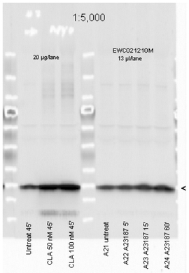
- Figure 2. Immunoblotting. Affinity Purified Phospho specific antibody to Monophosphorylated Regulatory Light Chain of Smooth and Non-muscle Myosin at pS19/pS20 was used at a 1:5000 dilution to detect myosin light chain by Western blot. Either 13 or 20 µl of a mouse cardiac myocyte lysate was loaded on a 4-20% Criterion gel for SDS-PAGE. Samples were either mocktreated or CLA-treated, as indicated. After washing, a 1:5,000 dilution of HRP conjugated Gt-a-Rabbit IgG preceded color development using Amersham's substrate system. Other detection methods will yield similar results. Data courtesy of the Alliance for Cellular Signaling (http://www.signaling-gateway.org).
- Submitted by
- Acris Antibodies GmbH (provider)
- Main image
- Experimental details
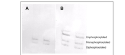
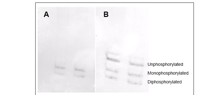
- Figure 3. Immunoblotting. Affinity purified phosphospecific antibody to phosphorylated regulatory light chain of smooth and non-muscle Myosin at pS19/pS20 was used at a 1:1000 dilution to detect myosin light chain by Western blot on 3T3 cell lysates. A standard urea/glycerol gel without SDS was used to separate phospho forms of regulatory light chain according to mass to charge ratios. In Panel A, reactivity of phosphospecific antibody is shown. In Panel B, reactivity of commercially available pan reactive antibody that detects both unphosphorylated and phosphorylated forms of regulatory light chain is shown. Phosphospecific antibody detects both monophosphorylated (pSer20 Mono-P-RLC) and diphosphorylated (pThr19-pSer20 Di-P-RLC) regulatory light chain. Personal communication. J. Stull. UT Southwestern Medical Center.
Supportive validation
- Submitted by
- Acris Antibodies GmbH (provider)
- Main image

- Experimental details
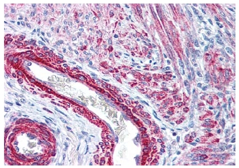
- Figure 1. Immunohistochemistry. Polyclonal anti-Monophosphorylated RLC Smooth and Non-Muscle Myosin pS19/20 antibody (R1535P) was used at 2.5 µg/ml to detect signal in a variety of tissues including multi-human, multi-brain and multi-cancer slides. This image shows strong staining of both vascular and myometrial smooth muscle cells of the uterus. Tissue was formalin-fixed and paraffin embedded. The image shows localization of the antibody as the precipitated red signal, with a hematoxylin purple nuclear counterstain. Personal Communication, Tina Roush, LifeSpanBiosciences, Seattle, WA.