Explore
 Validate
 Learn
Learn Western blot
Antibody data
- Antibody Data
- Antigen structure
- References [4]
- Comments [0]
- Validations
- Western blot [1]
- Immunohistochemistry [1]
Submit
Validation data
Reference
Comment
Report error
- Product number
- sc-8405 - Provider product page

- Provider
- Santa Cruz Biotechnology
- Proper citation
- Santa Cruz Biotechnology Cat#sc-8405, RRID:AB_628014
- Product name
- Anti-NFATC3
- Antibody type
- Monoclonal
- Antigen
- Recombinant full-length protein
- Reactivity
- Human
- Host
- Mouse
Submitted references Calcium/NFAT signalling promotes early nephrogenesis.
Peroxisome proliferator-activated receptor beta activation promotes myonuclear accretion in skeletal muscle of adult and aged mice.
Cholecystokinin activates pancreatic calcineurin-NFAT signaling in vitro and in vivo.
Calcineurin-GATA-6 pathway is involved in smooth muscle-specific transcription.
Burn SF, Webb A, Berry RL, Davies JA, Ferrer-Vaquer A, Hadjantonakis AK, Hastie ND, Hohenstein P
Developmental biology 2011 Apr 15;352(2):288-98
Developmental biology 2011 Apr 15;352(2):288-98
Peroxisome proliferator-activated receptor beta activation promotes myonuclear accretion in skeletal muscle of adult and aged mice.
Giordano C, Rousseau AS, Wagner N, Gaudel C, Murdaca J, Jehl-Piétri C, Sibille B, Grimaldi PA, Lopez P
Pflugers Archiv : European journal of physiology 2009 Sep;458(5):901-13
Pflugers Archiv : European journal of physiology 2009 Sep;458(5):901-13
Cholecystokinin activates pancreatic calcineurin-NFAT signaling in vitro and in vivo.
Gurda GT, Guo L, Lee SH, Molkentin JD, Williams JA
Molecular biology of the cell 2008 Jan;19(1):198-206
Molecular biology of the cell 2008 Jan;19(1):198-206
Calcineurin-GATA-6 pathway is involved in smooth muscle-specific transcription.
Wada H, Hasegawa K, Morimoto T, Kakita T, Yanazume T, Abe M, Sasayama S
The Journal of cell biology 2002 Mar 18;156(6):983-91
The Journal of cell biology 2002 Mar 18;156(6):983-91
No comments: Submit comment
Supportive validation
- Submitted by
- per
- Main image
- Experimental details
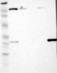
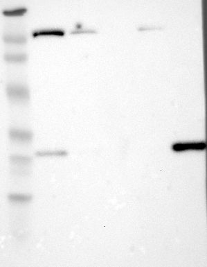
- Western blot analysis of antibody specificity using a routine panel composed of IgG/HSA-depleted human plasma and protein lysates from selected human tissues and cell lines.
- Validation comment
- Band of predicted size in kDa (+/-20%) with additional bands present.
- Primary Ab dilution
- 1:500
- Secondary Ab dilution
- 1:7000
- Lane 1
- Marker [kDa]: 230, 110, 82, 49.3, 32.2, 25.5, 17.6
- Lane 2
- RT-4
- Lane 3
- U-251MG sp
- Lane 4
- Human Plasma
- Lane 5
- Liver
- Lane 6
- Tonsil
- Theoretical target weight
- [kDa] 66
Supportive validation
- Submitted by
- per
- Main image

- Experimental details
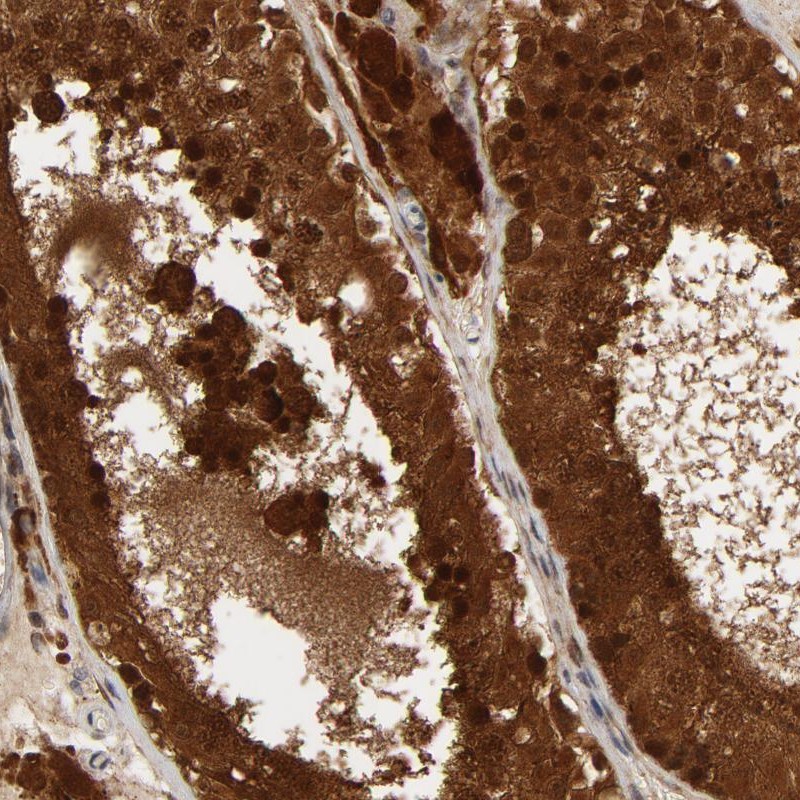
- Immunohistochemical staining of human testis shows strong cytoplasmic and nuclear positivity in cells in seminiferus ducts.
- Validation comment
- Staining pattern consistent with experimental and/or bioinformatic data.