Explore
 Validate
 Learn
Learn Western blot
 Immunohistochemistry
Antibody data
- Antibody Data
- Antigen structure
- References [4]
- Comments [0]
- Validations
- Western blot [1]
- Immunocytochemistry [2]
Submit
Validation data
Reference
Comment
Report error
- Product number
- HPA024120 - Provider product page

- Provider
- Atlas Antibodies
- Proper citation
- Atlas Antibodies Cat#HPA024120, RRID:AB_1858191
- Product name
- Anti-TOP2B
- Antibody type
- Polyclonal
- Description
- Polyclonal Antibody against Human TOP2B, Gene description: topoisomerase (DNA) II beta 180kDa, Validated applications: WB, IHC, ICC, Uniprot ID: Q02880, Storage: Store at +4°C for short term storage. Long time storage is recommended at -20°C.
- Reactivity
- Human, Mouse, Rat
- Host
- Rabbit
- Conjugate
- Unconjugated
- Isotype
- IgG
- Vial size
- 100 µl
- Concentration
- 0.4 mg/ml
- Storage
- Store at +4°C for short term storage. Long time storage is recommended at -20°C.
- Handling
- The antibody solution should be gently mixed before use.
Submitted references Discovery of novel DNA‐damaging agents through phenotypic screening for DNA double‐strand break
Genome-wide prediction of topoisomerase IIβ binding by architectural factors and chromatin accessibility
Histone H2A phosphorylation recruits topoisomerase II α to centromeres to safeguard genomic stability
Uncovering Hidden Layers of Cell Cycle Regulation through Integrative Multi-omic Analysis
Zhang D, Shimokawa T, Guo Q, Dan S, Miki Y, Sunada S
Cancer Science 2022;114(3):1108-1117
Cancer Science 2022;114(3):1108-1117
Genome-wide prediction of topoisomerase IIβ binding by architectural factors and chromatin accessibility
Ay F, Martínez-García P, García-Torres M, Divina F, Terrón-Bautista J, Delgado-Sainz I, Gómez-Vela F, Cortés-Ledesma F
PLOS Computational Biology 2021;17(1):e1007814
PLOS Computational Biology 2021;17(1):e1007814
Histone H2A phosphorylation recruits topoisomerase II α to centromeres to safeguard genomic stability
Zhang M, Liang C, Chen Q, Yan H, Xu J, Zhao H, Yuan X, Liu J, Lin S, Lu W, Wang F
The EMBO Journal 2019;39(3)
The EMBO Journal 2019;39(3)
Uncovering Hidden Layers of Cell Cycle Regulation through Integrative Multi-omic Analysis
Snyder M, Aviner R, Shenoy A, Elroy-Stein O, Geiger T
PLOS Genetics 2015;11(10):e1005554
PLOS Genetics 2015;11(10):e1005554
No comments: Submit comment
Enhanced validation
- Submitted by
- Atlas Antibodies (provider)
- Enhanced method
- Genetic validation
- Main image

- Experimental details
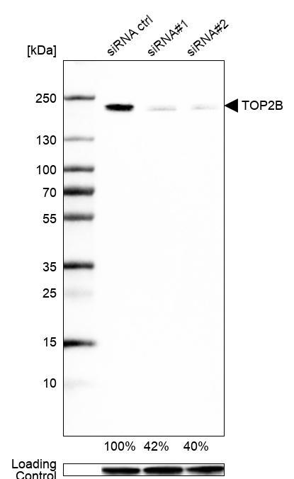
- Western blot analysis in U2OS cells transfected with control siRNA, target specific siRNA probe #1 and #2, using Anti-TOP2B antibody. Remaining relative intensity is presented. Loading control: Anti-GAPDH.
- Sample type
- Human
- Protocol
- Protocol
Enhanced validation
Supportive validation
- Submitted by
- 55af80e3e0991
- Enhanced method
- Genetic validation
- Main image
- Experimental details
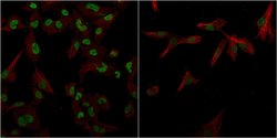
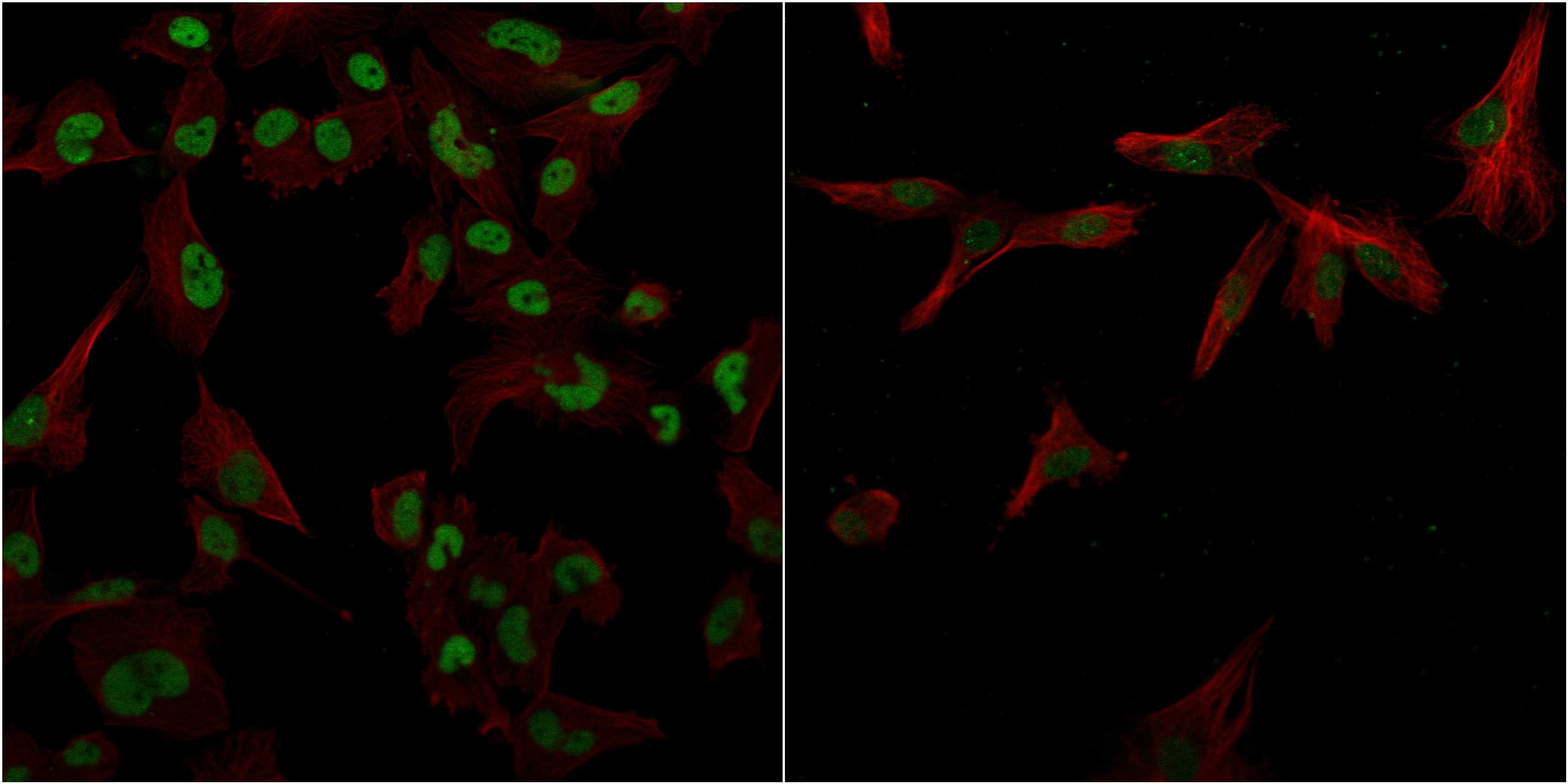
- Confocal images of immunofluorescently stained human U-2 OS cells.The protein TOP2B is shown in green and the microtubules in red. The image to the left show cells transfected with control siRNA and the image to the right show cells where TOP2B has been downregulated with specific siRNA.
- Sample type
- U-2 OS cells
- Primary Ab dilution
- 1:105
- Secondary Ab
- Secondary Ab
- Secondary Ab dilution
- 1:800
- Knockdown/Genetic Approaches Application
- Immunocytochemistry
Supportive validation
- Submitted by
- Atlas Antibodies (provider)
- Main image

- Experimental details
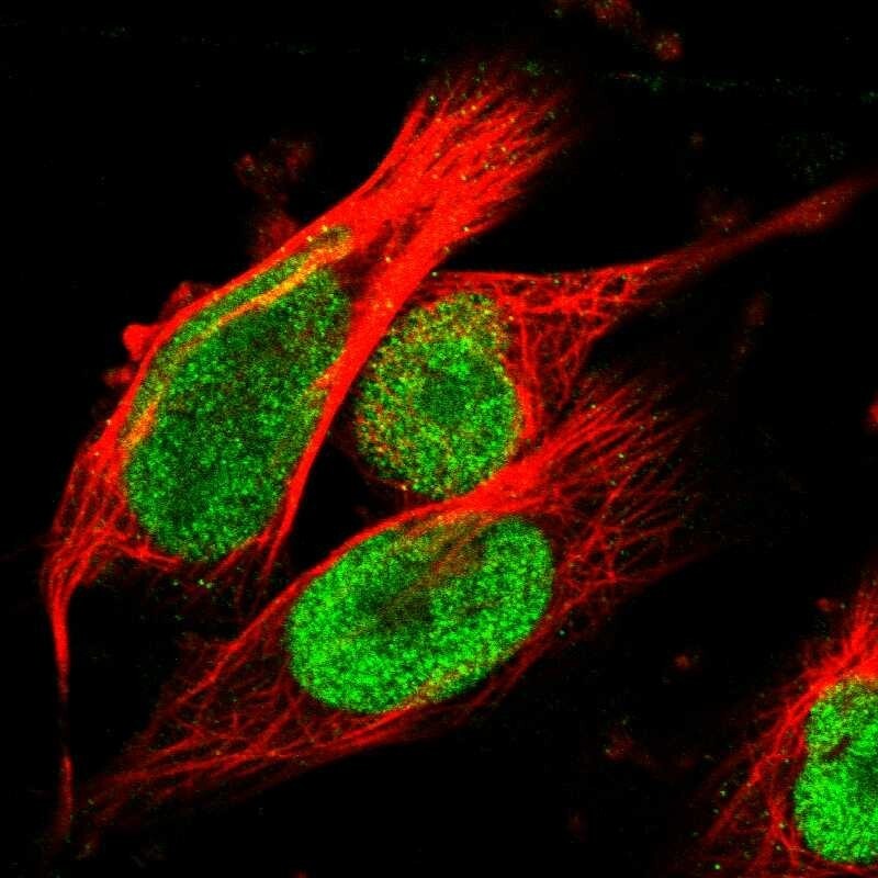
- Immunofluorescent staining of human cell line U-251 MG shows localization to nucleoplasm.
- Sample type
- Human