 Explore
 Validate
 Learn
Learn Western blot
Antibody data
- Antibody Data
- Antigen structure
- References [2]
- Comments [0]
- Validations
- Western blot [4]
- Immunocytochemistry [2]
- Immunoprecipitation [1]
- Immunohistochemistry [6]
- Other assay [4]
Submit
Validation data
Reference
Comment
Report error
- Product number
- PA5-29622 - Provider product page

- Provider
- Invitrogen Antibodies
- Product name
- TRAF6 Polyclonal Antibody
- Antibody type
- Polyclonal
- Antigen
- Recombinant full-length protein
- Description
- Recommended positive controls: A431, A549, HeLa, HepG2, NIH-3T3, BCL-1, PC-12, Rat2. Predicted reactivity: Mouse (80%), Pig (83%), Rhesus Monkey (97%), Bovine (82%). Store product as a concentrated solution. Centrifuge briefly prior to opening the vial.
- Reactivity
- Human, Mouse, Rat
- Host
- Rabbit
- Isotype
- IgG
- Vial size
- 100 μL
- Concentration
- 3.39 mg/mL
- Storage
- Store at 4°C short term. For long term storage, store at -20°C, avoiding freeze/thaw cycles.
Submitted references Myeloid Differentiation Primary Response 88-Cyclin D1 Signaling in Breast Cancer Cells Regulates Toll-Like Receptor 3-Mediated Cell Proliferation.
A truncating mutation in the autophagy gene UVRAG drives inflammation and tumorigenesis in mice.
Singh A, Devkar R, Basu A
Frontiers in oncology 2020;10:1780
Frontiers in oncology 2020;10:1780
A truncating mutation in the autophagy gene UVRAG drives inflammation and tumorigenesis in mice.
Quach C, Song Y, Guo H, Li S, Maazi H, Fung M, Sands N, O'Connell D, Restrepo-Vassalli S, Chai B, Nemecio D, Punj V, Akbari O, Idos GE, Mumenthaler SM, Wu N, Martin SE, Hagiya A, Hicks J, Cui H, Liang C
Nature communications 2019 Dec 12;10(1):5681
Nature communications 2019 Dec 12;10(1):5681
No comments: Submit comment
Supportive validation
- Submitted by
- Invitrogen Antibodies (provider)
- Main image
- Experimental details
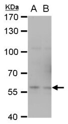
- TRAF6 Polyclonal Antibody detects TRAF6 protein by western blot analysis. A. 30 µg PC-12 whole cell extract. B. 30 µg Rat2 whole cell extract.7.5% SDS-PAGE. TRAF6 Polyclonal Antibody (Product # PA5-29622) dilution: 1:1,000. The HRP-conjugated anti-rabbit IgG antibody was used to detect the primary antibody.
- Submitted by
- Invitrogen Antibodies (provider)
- Main image

- Experimental details
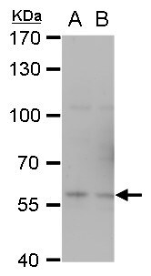
- Western Blot using TRAF6 Polyclonal Antibody (Product # PA5-29622). Sample (30 µg of whole cell lysate). Lane A: NIH-3T3. Lane B: BCL-1. 7.5% SDS PAGE. TRAF6 Polyclonal Antibody (Product # PA5-29622) diluted at 1:1,000. The HRP-conjugated anti-rabbit IgG antibody was used to detect the primary antibody.
- Submitted by
- Invitrogen Antibodies (provider)
- Main image
- Experimental details
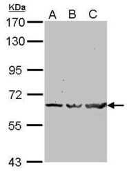
- Western Blot using TRAF6 Polyclonal Antibody (Product # PA5-29622). Sample (30 µg of whole cell lysate). Lane A: A549. Lane B: HeLa. Lane C: HepG2. 7.5% SDS PAGE. TRAF6 Polyclonal Antibody (Product # PA5-29622) diluted at 1:1,000. The HRP-conjugated anti-rabbit IgG antibody was used to detect the primary antibody.

- Submitted by
- Invitrogen Antibodies (provider)
- Main image
- Experimental details
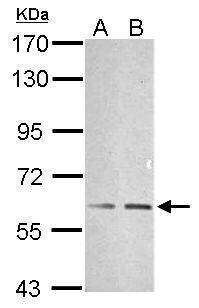
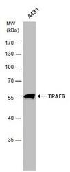
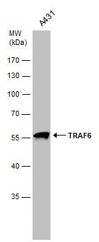
- Western Blot analysis of TRAF6 was performed by separating 30 µg of whole cell extract by 10% SDS-PAGE. Proteins were transferred to a membrane and probed with a TRAF6 Polyclonal Antibody (Product # PA5-29622) at a dilution of 1:2000.
Supportive validation
- Submitted by
- Invitrogen Antibodies (provider)
- Main image

- Experimental details
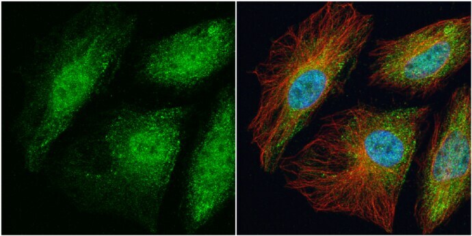
- Immunocytochemistry-Immunofluorescence analysis of TRAF6 was performed in HeLa cells fixed in 4% paraformaldehyde at RT for 15 min. Green: TRAF6 Polyclonal Antibody (Product # PA5-29622) diluted at 1:100. Red: alpha Tubulin, a cytoskeleton marker. Blue: Hoechst 33342 staining.
- Submitted by
- Invitrogen Antibodies (provider)
- Main image
- Experimental details
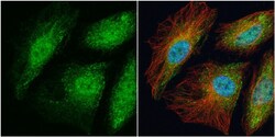
- Immunocytochemistry-Immunofluorescence analysis of TRAF6 was performed in HeLa cells fixed in 4% paraformaldehyde at RT for 15 min. Green: TRAF6 Polyclonal Antibody (Product # PA5-29622) diluted at 1:100. Red: alpha Tubulin, a cytoskeleton marker. Blue: Hoechst 33342 staining.

Supportive validation
- Submitted by
- Invitrogen Antibodies (provider)
- Main image

- Experimental details
- TRAF6 Polyclonal Antibody immunoprecipitates TRAF6 protein in IP experiments. IP samples: K562 whole cell extract. A. Control with 4 µg of preimmune Rabbit IgG. B. Immunoprecipitation of TRAF6 protein by 4 µg TRAF6 Polyclonal Antibody (Product # PA5-29622). 7.5 % SDS-PAGE. The immunoprecipitated TRAF6 protein was detected by TRAF6 Polyclonal Antibody (Product # PA5-29622) diluted at 1:500.

Supportive validation
- Submitted by
- Invitrogen Antibodies (provider)
- Main image

- Experimental details
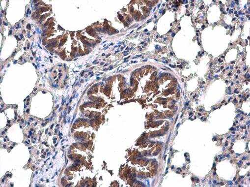
- Immunohistochemistry (Paraffin) analysis of TRAF6 was performed in paraffin-embedded mouse lung tissue using TRAF6 Polyclonal Antibody (Product # PA5-29622) at a dilution of 1:500.
- Submitted by
- Invitrogen Antibodies (provider)
- Main image
- Experimental details
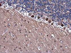
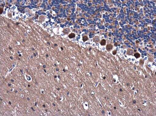
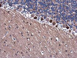
- Immunohistochemistry (Paraffin) analysis of TRAF6 was performed in paraffin-embedded rat brain tissue using TRAF6 Polyclonal Antibody (Product # PA5-29622) at a dilution of 1:500.
- Submitted by
- Invitrogen Antibodies (provider)
- Main image

- Experimental details
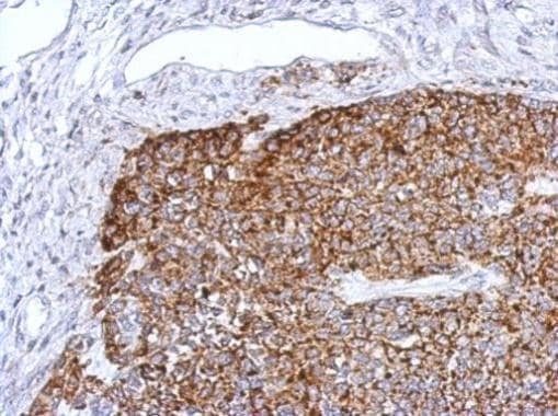
- Immunohistochemical analysis of paraffin-embedded 59T xenograft, using TRAF6 (Product # PA5-29622) antibody at 1:500 dilution. Antigen Retrieval: EDTA based buffer, pH 8.0, 15 min.

- Submitted by
- Invitrogen Antibodies (provider)
- Main image

- Experimental details
- Immunohistochemistry (Paraffin) analysis of TRAF6 was performed in paraffin-embedded mouse lung tissue using TRAF6 Polyclonal Antibody (Product # PA5-29622) at a dilution of 1:500.

- Submitted by
- Invitrogen Antibodies (provider)
- Main image
- Experimental details
- Immunohistochemistry (Paraffin) analysis of TRAF6 was performed in paraffin-embedded rat brain tissue using TRAF6 Polyclonal Antibody (Product # PA5-29622) at a dilution of 1:500.

- Submitted by
- Invitrogen Antibodies (provider)
- Main image

- Experimental details
- Immunohistochemical analysis of paraffin-embedded 59T xenograft, using TRAF6 (Product # PA5-29622) antibody at 1:500 dilution. Antigen Retrieval: EDTA based buffer, pH 8.0, 15 min.
Supportive validation
- Submitted by
- Invitrogen Antibodies (provider)
- Main image

- Experimental details
- TRAF6 Polyclonal Antibody immunoprecipitates TRAF6 protein in IP experiments. IP samples: K562 whole cell extract. A. Control with 4 µg of preimmune Rabbit IgG. B. Immunoprecipitation of TRAF6 protein by 4 µg TRAF6 Polyclonal Antibody (Product # PA5-29622). 7.5 % SDS-PAGE. The immunoprecipitated TRAF6 protein was detected by TRAF6 Polyclonal Antibody (Product # PA5-29622) diluted at 1:500.

- Submitted by
- Invitrogen Antibodies (provider)
- Main image
- Experimental details
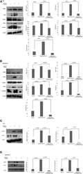
- Figure 5 Western blotting for the expression of signaling protein. (A,B) Cell lysate were collected and subjected to Western blot assay to estimate the level of expression of interleukin 1 receptor-associated kinase 1 (IRAK1), transforming growth factor beta-activated kinase 1 (TAK1), TGF-beta-activated kinase 1 (TAB1), TNF receptor-associated factor 6 (TRAF6), and cyclin D1. (C,D) Expression of pIRAK1 and pTAK1. beta-actin was used as loading control. The respective bar graphs are presented as densitometry analysis as mean +- SD of experiments ( p < 0.05 is treated as significant).

- Submitted by
- Invitrogen Antibodies (provider)
- Main image
- Experimental details
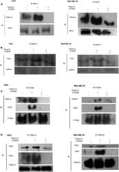
- Figure 6 Immunoprecipitation showing the involvement of the signaling complex. (A) Signaling complex of IRAK1/TRAF6 was immunoprecipitated with antibodies against IRAK1 followed by Western blotting with anti-TRAF6 and anti-IRAK1 antibodies. (B) Signaling complex of pIRAK1/TAK1 was immunoprecipitated with antibodies against pIRAK1 followed by Western blotting with anti-TAK1 and anti-pIRAK1 antibodies. (C) Signaling complex TAB1-TRAF6-TAK1 was immunoprecipitated with antibodies against pTAK1 followed by Western blotting using anti-TRAF6, TAB1, and pTAK1 antibodies. (D) Signaling complex TAB1-TRAF6-TAK1 was immunoprecipitated with antibodies against TRAF6 followed by Western blotting analysis using anti-TAK1, TAB1, and TRAF6 antibodies.

- Submitted by
- Invitrogen Antibodies (provider)
- Main image
- Experimental details
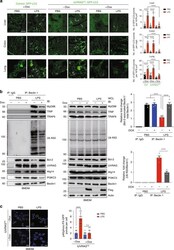
- Fig. 2 UVRAG FS prevents TLR4 induction of autophagy in vivo. a Representative images (left panels) and quantification (right panels) of GFP-LC3 puncta in indicated tissues from Dox-treated/untreated iUVRAG FS mice and littermate control mice crossed with GFP-LC3 transgenic mice, challenged with PBS or LPS (20 mg per kg body weight; i.p.). Arrows denote GFP-LC3 puncta. Scale bars, 10 mum. n = 10 tissue sections pooled from three independent experiments. b UVRAG FS inhibits the Beclin1-autophagy interactome and K63-linked ubiquitination in LPS-stimulated bone marrow-derived macrophages (BMDMs). BMDMs derived from iUVRAG FS mice were incubated with PBS or LPS (100 ng/mL) for 4 h in the presence/absence of Dox and subjected to IP with anti-Beclin1 followed by WB of indicated proteins (left panel). Expression of indicated proteins in WCL were shown (middle panel). Densitometric quantification of the Bcl-2-associated Beclin1/total Beclin1 and the ubiquitinated (K63)-Beclin1/total Beclin1 ratios in BMDM are also shown (right panels). c UVRAG FS prevents LPS-induced PI3KC3 activation in BMDM. Cells cultured as in b were transfected with p40(phox)-PX-GFP and subjected to confocal microscopy. Representative images of p40(phox)-PX-GFP puncta in indicated BMDM are shown (left) and the number of p40(phox)-PX-GFP puncta per cell was quantified (right). Scale bars, 10 mum. n = 10-15 high-power fields (HPF) pooled from three independent experiments. Data in b are from one experiment that is
