Explore
 Validate
 Learn
Learn Western blot
Antibody data
- Antibody Data
- Antigen structure
- References [0]
- Comments [0]
- Validations
- Western blot [1]
- Immunocytochemistry [1]
- Immunohistochemistry [2]
- Flow cytometry [1]
Submit
Validation data
Reference
Comment
Report error
- Product number
- ANT-018-200UL - Provider product page

- Provider
- Invitrogen Antibodies
- Product name
- TrkA (extracellular) Polyclonal Antibody
- Antibody type
- Polyclonal
- Antigen
- Other
- Reactivity
- Human, Mouse, Rat
- Host
- Rabbit
- Isotype
- IgG
- Vial size
- 200 µL
- Concentration
- 0.8 mg/mL
- Storage
- -20° C, Avoid Freeze/Thaw Cycles
No comments: Submit comment
Supportive validation
- Submitted by
- Invitrogen Antibodies (provider)
- Main image
- Experimental details
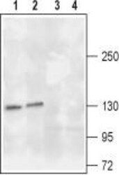
- Western blot analysis of rat (lanes 1 and 3) and mouse (lanes 2 and 4) brain lysates: - 1,2. Anti-TrkA (extracellular) Antibody (#ANT-018), (1:200).3,4. Anti-TrkA (extracellular) Antibody , preincubated with TrkA (extracellular) Blocking Peptide (#BLP-NT018).

Supportive validation
- Submitted by
- Invitrogen Antibodies (provider)
- Main image

- Experimental details
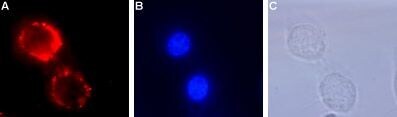
- Expression of TrkA in live intact rat PC12 cells - Cell surface detection of TrkA in live intact rat PC12 cells. A. Cells were stained with Anti-TrkA (extracellular) Antibody (#ANT-018) (1:50), followed by goat Anti-rabbit-AlexaFluor-494 secondary Antibody (red). B. Cell nuclei were visualized with the membrane-permeable DNA dye Hoechst 33342 (blue staining). C. Live view of the cells.
Supportive validation
- Submitted by
- Invitrogen Antibodies (provider)
- Main image
- Experimental details
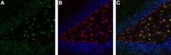
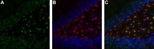
- Expression of TrkA in rat brain hippocampal dentate gyrus - Immunohistochemical staining of immersion-fixed, free floating rat brain frozen sections. A. Brain sections were stained using Anti-TrkA (extracellular) Antibody (#ANT-018), (1:1000), (green staining). B. The same section was also stained for glial fibrillary acidic protein (GFAP) (red and counterstained blue).C. Overlay of A and B demonstrates co-localization of TrkA and GFAP in dentate gyrus astrocytes.
- Submitted by
- Invitrogen Antibodies (provider)
- Main image
- Experimental details
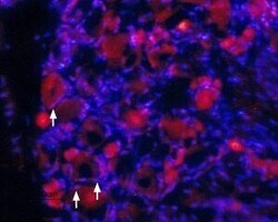
- Expression of TrkA in rat DRG - Immunohistochemical staining of rat dorsal root ganglia (DRG) frozen sections using Anti-TrkA (extracellular) Antibody (#ANT-018), (1:100). TrkA (red staining) is expressed in DRG neurons and in satellite microglia (arrows). Hoechst 33342 is used as the counterstain.

Supportive validation
- Submitted by
- Invitrogen Antibodies (provider)
- Main image

- Experimental details
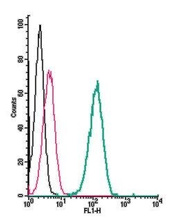
- Cell surface detection of TrkA by indirect flow cytometry in live intact human THP-1 monocytic leukemia cells: - (black line) cells. (red) Cells + goat- Anti-rabbit-FITC. (green) Cells + Anti-TrkA (extracellular) Antibody (#ANT-018), (2.5μg) + goat- Anti-rabbit-FITC.