 Explore
 Validate
 Learn
Learn Immunohistochemistry
Antibody data
- Antibody Data
- Antigen structure
- References [0]
- Comments [0]
- Validations
- Immunohistochemistry [2]
Submit
Validation data
Reference
Comment
Report error
- Product number
- ANT-018-FR-50UL - Provider product page

- Provider
- Invitrogen Antibodies
- Product name
- TrkA (extracellular) Polyclonal Antibody, Atto 633
- Antibody type
- Polyclonal
- Antigen
- Other
- Reactivity
- Human, Mouse, Rat
- Host
- Rabbit
- Conjugate
- Red dye
- Isotype
- IgG
- Vial size
- 50 µL
- Concentration
- 1 mg/mL
- Storage
- -20° C, Avoid Freeze/Thaw Cycles
No comments: Submit comment
Supportive validation
- Submitted by
- Invitrogen Antibodies (provider)
- Main image
- Experimental details
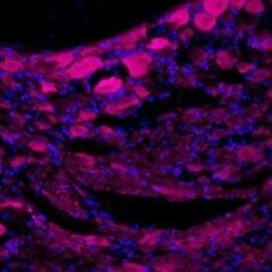
- Expression of TrkA in rat dorsal root ganglion - Immunohistochemical staining of rat dorsal root ganglia (DRG) frozen sections using Anti-TrkA (extracellular)-ATTO Fluor-633 Antibody (#ANT-018-FR), (1:50). TrkA (purple staining) is expressed in DRG neurons. Hoechst 33342 (blue) is used as the counterstain.

- Submitted by
- Invitrogen Antibodies (provider)
- Main image
- Experimental details
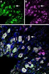
- Immunolocalization of p75NTRand TrkA in rat dorsal root ganglion - Immunohistochemical staining of perfusion-fixed frozen rat dorsal root ganglia (DRG) sections using Anti-p75 NGF Receptor (extracellular)-ATTO Fluor-488 Antibody (#ANT-007-AG), (1:60) and Anti-TrkA (extracellular)-ATTO Fluor-633 Antibody (#ANT-018-FR), (1:60). A. Sections were labeled with Anti-p75 NGR Receptor (extracellular)-ATTO Fluor-488 Antibody (green). B. The same section was stained with Anti-TrkA (extracellular)-ATTO Fluor-633 Antibody (purple). C. Merge of the two images suggests extensive co-localization (arrow points at an example) of TrkA and p75NTR receptors in rat dorsal root ganglion. Cell nuclei were stained with DAPI (blue).
