 Explore
 Validate
 Learn
Learn Western blot
Antibody data
- Antibody Data
- Antigen structure
- References [0]
- Comments [0]
- Validations
- Western blot [5]
- Immunocytochemistry [2]
- Immunohistochemistry [3]
Submit
Validation data
Reference
Comment
Report error
- Product number
- PA5-28886 - Provider product page

- Provider
- Invitrogen Antibodies
- Product name
- TrxR1 Polyclonal Antibody
- Antibody type
- Polyclonal
- Antigen
- Recombinant protein fragment
- Description
- Recommended positive controls: Molt-4, mouse brain, rat brain.
- Concentration
- 1 mg/mL
No comments: Submit comment
Supportive validation
- Submitted by
- Invitrogen Antibodies (provider)
- Main image
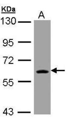
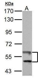
- Experimental details
- Western Blot using TrxR1 Polyclonal Antibody (Product # PA5-28886). Sample (30 µg of whole cell lysate). A: Molt-4. 7.5% SDS PAGE. TrxR1 Polyclonal Antibody (Product # PA5-28886) diluted at 1:3,000. The HRP-conjugated anti-rabbit IgG antibody was used to detect the primary antibody.

- Submitted by
- Invitrogen Antibodies (provider)
- Main image
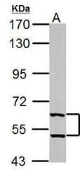
- Experimental details
- TrxR1 Polyclonal Antibody detects TXNRD1 protein by western blot analysis. A. 50 µg mouse brain lysate/extract. 7.5% SDS-PAGE. TrxR1 Polyclonal Antibody (Product # PA5-28886) dilution: 1:1,000. The HRP-conjugated anti-rabbit IgG antibody was used to detect the primary antibody.
- Submitted by
- Invitrogen Antibodies (provider)
- Main image
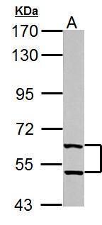
- Experimental details
- TrxR1 Polyclonal Antibody detects TXNRD1 protein by western blot analysis. A. 50 µg Rat brain lysate/extract. 7.5% SDS-PAGE. TrxR1 Polyclonal Antibody (Product # PA5-28886) dilution: 1:1,000. The HRP-conjugated anti-rabbit IgG antibody was used to detect the primary antibody.
- Submitted by
- Invitrogen Antibodies (provider)
- Main image
- Experimental details
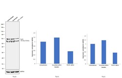
- Knockdown of TrxR1 was achieved by transfecting A549 with TrxR1 specific siRNAs (Silencer® select Product # S756, S757). Western blot analysis (Fig. a) was performed using Whole cell extracts from the TrxR1 knockdown cells (lane 3), non-targeting scrambled siRNA transfected cells (lane 2) and untransfected cells (lane 1). The blot was probed with TrxR1 Polyclonal Antibody (Product # PA5-28886, 1:1000 dilution ) and Goat anti-Rabbit IgG (H+L) Superclonal™ Recombinant Secondary Antibody, HRP (Product # A27036, 1:4000 dilution). Densitometric analysis of this western blot is shown in histogram (Fig. b) corresponding to lower band and histogram in Fig (c) corresponding to upper band show Decrease in signal upon siRNA mediated knock down confirms that antibody is specific to TrxR1.

- Submitted by
- Invitrogen Antibodies (provider)
- Main image

- Experimental details
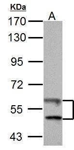
- Western blot was performed using Anti-TrxR1 Polyclonal Antibody (Product # PA5-28886) and a 55 kDa and 60 kDa band corresponding to TrxR1 was observed across cell lines. Whole cell extracts (30µg lysate) of A549 (Lane 1), HeLa (Lane 2), PC-3 (Lane 3) and MCF7 (Lane 4) were electrophoresed using NuPAGE™ 4-12% Bis-Tris Protein Gel (Product # NP0321BOX). Resolved proteins were then transferred onto a Nitrocellulose membrane (Product # IB23001) by iBlot® 2 Dry Blotting System (Product # IB21001). The blot was probed with the primary antibody (1:1000 dilution) and detected by chemiluminescence with Goat anti-Rabbit IgG (H+L) Superclonal™ Recombinant Secondary Antibody, HRP (Product # A27036, 1:4000 dilution) using the iBright FL 1000 (Product # A32752). Chemiluminescent detection was performed using Novex® ECL Chemiluminescent Substrate Reagent Kit (Product # WP20005).

Supportive validation
- Submitted by
- Invitrogen Antibodies (provider)
- Main image
- Experimental details
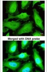
- Immunofluorescent analysis of TrxR1 in paraformaldehyde-fixed HeLa cells using a TrxR1 polyclonal antibody (Product # PA5-28886) at a 1:200 dilution.

- Submitted by
- Invitrogen Antibodies (provider)
- Main image
- Experimental details
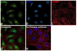
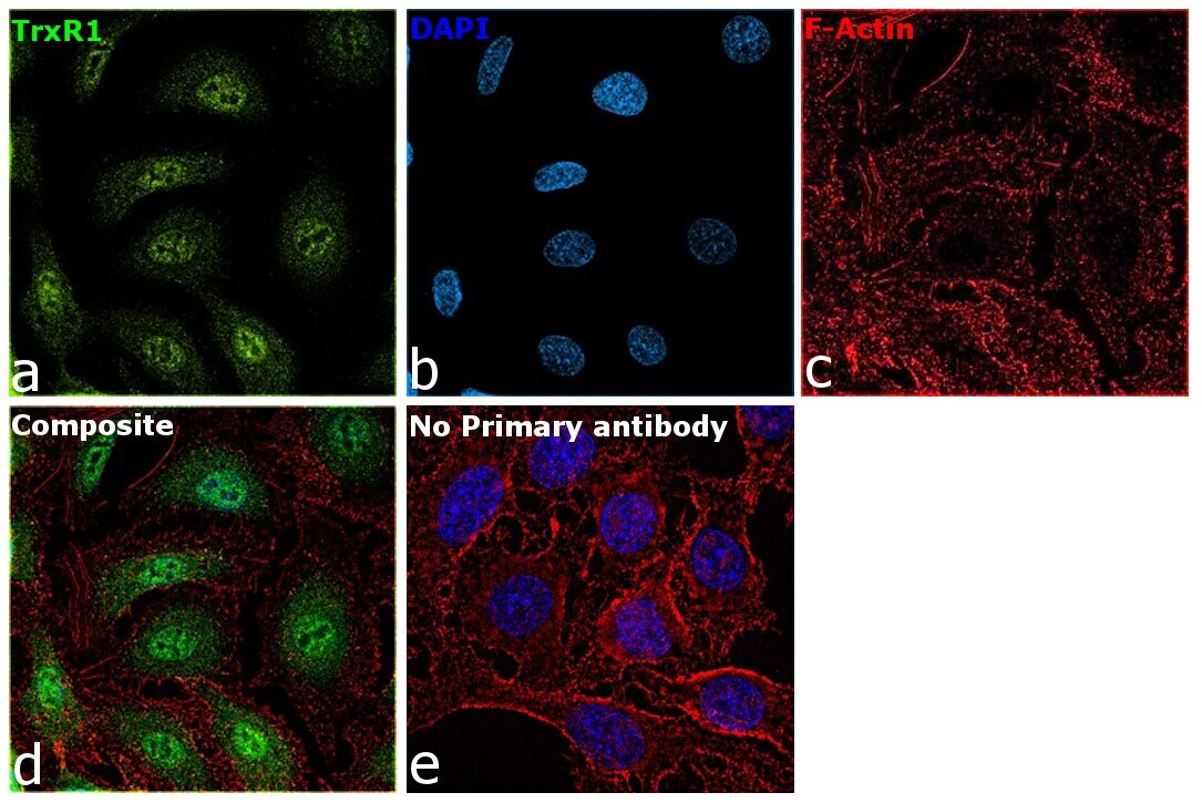
- Immunofluorescence analysis of TrxR1 was performed using 70 confluent log phase A549 cells. The cells were fixed with 4% paraformaldehyde for 10 minutes, permeabilized with 0.1% Triton™ X-100 for 15 minutes, and blocked with 2% BSA for 45 minutes at room temperature. The cells were labeled with TrxR1 Polyclonal Antibody (Product # PA5-28886) at 1:100 dilution in 0.1% BSA, incubated at 4 degree celsius overnight and then labeled with Donkey anti-Rabbit IgG (H+L) Highly Cross-Adsorbed Secondary Antibody, Alexa Fluor Plus 488 (Product # A32790), (1:2000 dilution), for 45 minutes at room temperature (Panel a: Green). Nuclei (Panel b:Blue) were stained with ProLong™ Diamond Antifade Mountant with DAPI (Product # P36962). F-actin (Panel c: Red) was stained with Rhodamine Phalloidin (Product # R415, 1:300 dilution). Panel d represents the merged image showing Nuclear and cytoplasmic localization. Panel e represents control cells with no primary antibody to assess background. The images were captured at 60X magnification.
Supportive validation
- Submitted by
- Invitrogen Antibodies (provider)
- Main image
- Experimental details
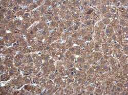
- Immunohistochemistry (Paraffin) analysis of TrxR1 was performed in paraffin-embedded mouse liver tissue using TrxR1 Polyclonal Antibody (Product # PA5-28886) at a dilution of 1:500.
- Submitted by
- Invitrogen Antibodies (provider)
- Main image
- Experimental details
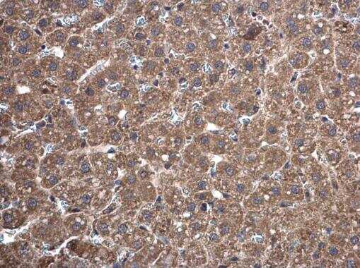
- Immunohistochemistry (Paraffin) analysis of TrxR1 was performed in paraffin-embedded mouse pancreas tissue using TrxR1 Polyclonal Antibody (Product # PA5-28886) at a dilution of 1:500.

- Submitted by
- Invitrogen Antibodies (provider)
- Main image
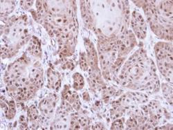
- Experimental details
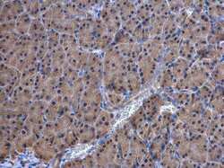
- Immunohistochemical analysis of paraffin-embedded Cal27 xenograft, using TrxR1 (Product # PA5-28886) antibody at 1:500 dilution. Antigen Retrieval: Citrate buffer, pH 6.0, 15 min.
