Explore
 Validate
 Learn
Learn Western blot
Antibody data
- Antibody Data
- Antigen structure
- References [1]
- Comments [0]
- Validations
- Western blot [1]
- Flow cytometry [1]
Submit
Validation data
Reference
Comment
Report error
- Product number
- MAB7428 - Provider product page

- Provider
- R&D Systems
- Product name
- Human/Mouse/Rat Thioredoxin Reductase 1/TRXR1 Antibody
- Antibody type
- Monoclonal
- Description
- Protein A or G purified from hybridoma culture supernatant. Detects mouse Thioredoxin Reductase 1/TRXR1 in direct ELISAs and human, mouse, and rat Thioredoxin Reductase 1/TRXR1 in Western blots.
- Reactivity
- Human, Mouse, Rat
- Host
- Mouse
- Conjugate
- Unconjugated
- Antigen sequence
Q16881- Isotype
- IgG
- Antibody clone number
- 489804
- Vial size
- 100 ug
- Concentration
- LYOPH
- Storage
- Use a manual defrost freezer and avoid repeated freeze-thaw cycles. 12 months from date of receipt, -20 to -70 °C as supplied. 1 month, 2 to 8 °C under sterile conditions after reconstitution. 6 months, -20 to -70 °C under sterile conditions after reconstitution.
Submitted references Monitoring thioredoxin redox with a genetically encoded red fluorescent biosensor.
Fan Y, Makar M, Wang MX, Ai HW
Nature chemical biology 2017 Sep;13(9):1045-1052
Nature chemical biology 2017 Sep;13(9):1045-1052
No comments: Submit comment
Supportive validation
- Submitted by
- R&D Systems (provider)
- Main image
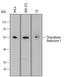
- Experimental details
- Detection of Human, Mouse, and Rat Thioredoxin Reductase 1/TRXR1 by Western Blot. Western blot shows lysates of HeLa human cervical epithelial carcinoma cell line, NIH-3T3 mouse embryonic fibroblast cell line, and C6 rat glioma cell line. PVDF membrane was probed with 0.1 µg/mL of Mouse Anti-Human Thioredoxin Reductase 1/TRXR1 Monoclonal Antibody (Catalog # MAB7428) followed by HRP-conjugated Anti-Mouse IgG Secondary Antibody (Catalog # HAF007). A specific band was detected for Thioredoxin Reductase 1/TRXR1 at approximately 65 kDa (as indicated). This experiment was conducted under reducing conditions and using Immunoblot Buffer Group 1.

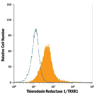
Supportive validation
- Submitted by
- R&D Systems (provider)
- Main image
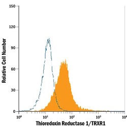
- Experimental details
- Detection of Thioredoxin Reductase 1/TRXR1 in HeLa Human Cell Line by Flow Cytometry. HeLa human cervical epithelial carcinoma cell line was stained with Mouse Anti-Human/Mouse/Rat Thioredoxin Reductase 1/TRXR1 Monoclonal Antibody (Catalog # MAB7428, filled histogram) or isotype control antibody (Catalog # MAB002, open histogram), followed by Allophycocyanin-conjugated Anti-Mouse IgG Secondary Antibody (Catalog # F0101B). To facilitate intracellular staining, cells were fixed with paraformaldehyde and permeabilized with saponin.