Explore
 Validate
 Learn
Learn Western blot
Antibody data
- Antibody Data
- Antigen structure
- References [4]
- Comments [0]
- Validations
- Western blot [1]
- Immunohistochemistry [1]
- Other assay [1]
Submit
Validation data
Reference
Comment
Report error
- Product number
- PAB0632 - Provider product page

- Provider
- Abnova Corporation
- Proper citation
- Abnova Corporation Cat#PAB0632, RRID:AB_983015
- Product name
- WEE1 (phospho S123) polyclonal antibody
- Antibody type
- Polyclonal
- Description
- Rabbit polyclonal antibody raised against synthetic phosphopeptide of WEE1.
- Storage
- Store at 4°C. For long term storage store at -20°C.Aliquot to avoid repeated freezing and thawing.
Submitted references Human Cdc14A regulates Wee1 stability by counteracting CDK-mediated phosphorylation.
M-phase kinases induce phospho-dependent ubiquitination of somatic Wee1 by SCFbeta-TrCP.
Keratinocyte G2/M growth arrest by 1,25-dihydroxyvitamin D3 is caused by Cdc2 phosphorylation through Wee1 and Myt1 regulation.
The clinical significance of Cyclin B1 and Wee1 expression in non-small-cell lung cancer.
Ovejero S, Ayala P, Bueno A, Sacristán MP
Molecular biology of the cell 2012 Dec;23(23):4515-25
Molecular biology of the cell 2012 Dec;23(23):4515-25
M-phase kinases induce phospho-dependent ubiquitination of somatic Wee1 by SCFbeta-TrCP.
Watanabe N, Arai H, Nishihara Y, Taniguchi M, Watanabe N, Hunter T, Osada H
Proceedings of the National Academy of Sciences of the United States of America 2004 Mar 30;101(13):4419-24
Proceedings of the National Academy of Sciences of the United States of America 2004 Mar 30;101(13):4419-24
Keratinocyte G2/M growth arrest by 1,25-dihydroxyvitamin D3 is caused by Cdc2 phosphorylation through Wee1 and Myt1 regulation.
Dai X, Yamasaki K, Yang L, Sayama K, Shirakata Y, Tokumara S, Yahata Y, Tohyama M, Hashimoto K
The Journal of investigative dermatology 2004 Jun;122(6):1356-64
The Journal of investigative dermatology 2004 Jun;122(6):1356-64
The clinical significance of Cyclin B1 and Wee1 expression in non-small-cell lung cancer.
Yoshida T, Tanaka S, Mogi A, Shitara Y, Kuwano H
Annals of oncology : official journal of the European Society for Medical Oncology / ESMO 2004 Feb;15(2):252-6
Annals of oncology : official journal of the European Society for Medical Oncology / ESMO 2004 Feb;15(2):252-6
No comments: Submit comment
Supportive validation
- Submitted by
- Abnova Corporation (provider)
- Main image
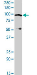
- Experimental details
- WEE1 (phospho S123) polyclonal antibody (Cat # PAB0632). Western blot analysis of WEE1 expression in human kidney.

Supportive validation
- Submitted by
- Abnova Corporation (provider)
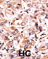
- Main image

- Experimental details
- Formalin-fixed and paraffin-embedded human hepatocellular carcinoma tissue reacted with WEE1 (phospho S123) polyclonal antibody (Cat # PAB0632) which was peroxidase-conjugated to the secondary antibody followed by AEC staining. This data demonstrates the use of this antibody for immunohistochemistry; clinical relevance has not been evaluated. HC = hepatocarcinoma
- Validation comment
- Immunohistochemistry (Formalin/PFA-fixed paraffin-embedded sections)
Supportive validation
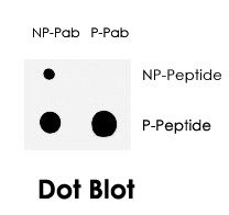
- Submitted by
- Abnova Corporation (provider)
- Main image
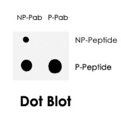
- Experimental details
- Dot blot analysis of WEE1 (phospho S123) polyclonal antibody (Cat # PAB0632) on nitrocellulose membrane. 50 ng of Phospho-peptide or Non Phospho-peptide per dot were adsorbed. Antobodies working concentration was 0.5&mciro;g/mL.
- Validation comment
- Dot Blot (Peptide)