Explore
 Validate
 Learn
Learn Western blot
 ELISA
ELISAAntibody data
- Antibody Data
- Antigen structure
- References [4]
- Comments [0]
- Validations
- Western blot [1]
- Immunohistochemistry [1]
- Other assay [1]
Submit
Validation data
Reference
Comment
Report error
- Product number
- PAB0633 - Provider product page

- Provider
- Abnova Corporation
- Proper citation
- Abnova Corporation Cat#PAB0633, RRID:AB_983016
- Product name
- WEE1 (phospho S53) polyclonal antibody
- Antibody type
- Polyclonal
- Antigen
- Synthetic phosphopeptide (conjugated with KLH) corresponding to residues surrounding S53 of human WEE1.
- Reactivity
- Human
- Host
- Rabbit
- Vial size
- 400 µl
- Storage
- Store at 4°C. For long term storage store at -20°C.Aliquot to avoid repeated freezing and thawing.
Submitted references Pim1 kinase is upregulated in glioblastoma multiforme and mediates tumor cell survival.
M-phase kinases induce phospho-dependent ubiquitination of somatic Wee1 by SCFbeta-TrCP.
Keratinocyte G2/M growth arrest by 1,25-dihydroxyvitamin D3 is caused by Cdc2 phosphorylation through Wee1 and Myt1 regulation.
The clinical significance of Cyclin B1 and Wee1 expression in non-small-cell lung cancer.
Herzog S, Fink MA, Weitmann K, Friedel C, Hadlich S, Langner S, Kindermann K, Holm T, Böhm A, Eskilsson E, Miletic H, Hildner M, Fritsch M, Vogelgesang S, Havemann C, Ritter CA, Meyer zu Schwabedissen HE, Rauch B, Hoffmann W, Kroemer HK, Schroeder H, Bien-Möller S
Neuro-oncology 2015 Feb;17(2):223-42
Neuro-oncology 2015 Feb;17(2):223-42
M-phase kinases induce phospho-dependent ubiquitination of somatic Wee1 by SCFbeta-TrCP.
Watanabe N, Arai H, Nishihara Y, Taniguchi M, Watanabe N, Hunter T, Osada H
Proceedings of the National Academy of Sciences of the United States of America 2004 Mar 30;101(13):4419-24
Proceedings of the National Academy of Sciences of the United States of America 2004 Mar 30;101(13):4419-24
Keratinocyte G2/M growth arrest by 1,25-dihydroxyvitamin D3 is caused by Cdc2 phosphorylation through Wee1 and Myt1 regulation.
Dai X, Yamasaki K, Yang L, Sayama K, Shirakata Y, Tokumara S, Yahata Y, Tohyama M, Hashimoto K
The Journal of investigative dermatology 2004 Jun;122(6):1356-64
The Journal of investigative dermatology 2004 Jun;122(6):1356-64
The clinical significance of Cyclin B1 and Wee1 expression in non-small-cell lung cancer.
Yoshida T, Tanaka S, Mogi A, Shitara Y, Kuwano H
Annals of oncology : official journal of the European Society for Medical Oncology / ESMO 2004 Feb;15(2):252-6
Annals of oncology : official journal of the European Society for Medical Oncology / ESMO 2004 Feb;15(2):252-6
No comments: Submit comment
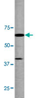
Supportive validation
- Submitted by
- Abnova Corporation (provider)
- Main image
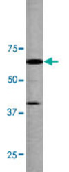
- Experimental details
- Western blot analysis of mouse liver tissue lysate with WEE1 (phospho S53) polyclonal antibody (Cat # PAB0633).
- Validation comment
- Western Blot (Tissue lysate)
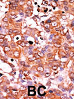
Supportive validation
- Submitted by
- Abnova Corporation (provider)
- Main image

- Experimental details
- Formalin-fixed and paraffin-embedded human breast carcinomareacted with WEE1 (phospho S53) polyclonal antibody (Cat # PAB0633), which was peroxidase-conjugated to the secondary antibody, followed by AEC staining.This data demonstrates the use of this antibody for immunohistochemistry; clinical relevance has not been evaluated.
- Validation comment
- Immunohistochemistry (Formalin/PFA-fixed paraffin-embedded sections)
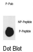
Supportive validation
- Submitted by
- Abnova Corporation (provider)
- Main image

- Experimental details
- Dot blot analysis of WEE1 (phospho S53) polyclonal antibody (Cat # PAB0633) on nitrocellulose membrane. 50ng of Phospho-peptide or Non Phospho-peptide per dot were adsorbed. Antibody working concentrations are 0.5ug per ml.
- Validation comment
- Dot Blot (Peptide)