Explore
 Validate
 Learn
Learn Western blot
Antibody data
- Antibody Data
- Antigen structure
- References [0]
- Comments [0]
- Validations
- Western blot [2]
- Immunocytochemistry [2]
Submit
Validation data
Reference
Comment
Report error
- Product number
- PA5-67019 - Provider product page

- Provider
- Invitrogen Antibodies
- Product name
- WEE1 Polyclonal Antibody
- Antibody type
- Polyclonal
- Antigen
- Recombinant full-length protein
- Description
- Immunogen sequence: LLKVMIHPDPE RRPSAMALVK HSVLLSASRK SAEQLRIELN AEKFKNSLLQ KELKKAQMAK AAAEERALFT DRMATRSTTQ Highest antigen sequence identity to the following orthologs - mouse 95%, rat 95%.
- Reactivity
- Human
- Host
- Rabbit
- Isotype
- IgG
- Vial size
- 100 µL
- Concentration
- 0.10 mg/mL
- Storage
- Store at 4°C short term. For long term storage, store at -20°C, avoiding freeze/thaw cycles.
No comments: Submit comment
Supportive validation
- Submitted by
- Invitrogen Antibodies (provider)
- Main image

- Experimental details
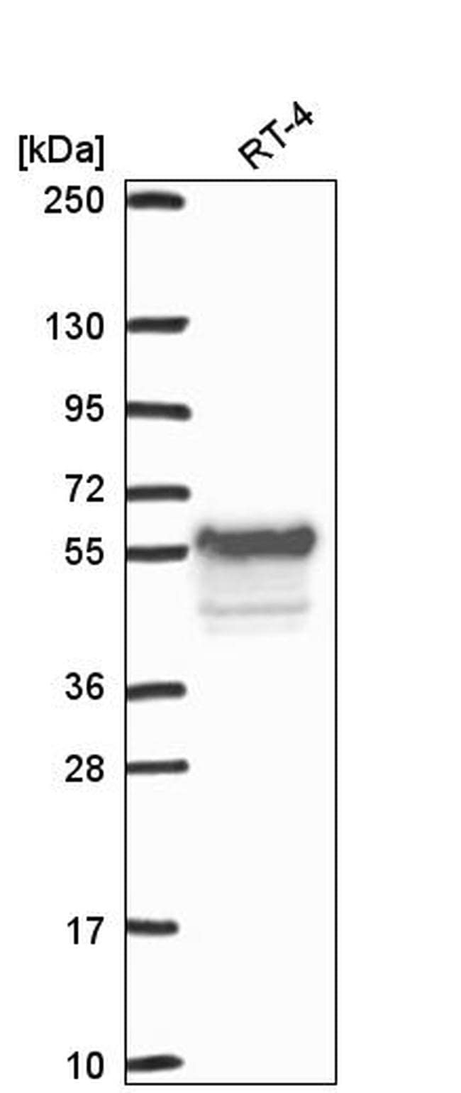
- Western blot analysis of WEE1 in human cell line RT-4. Samples were probed using a WEE1 Polyclonal Antibody (Product # PA5-67019).
- Submitted by
- Invitrogen Antibodies (provider)
- Main image
- Experimental details
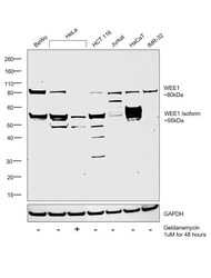
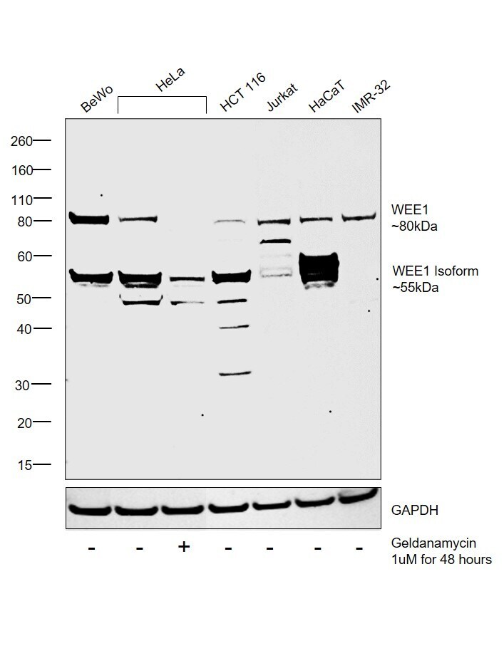
- Western blot was performed using Anti-WEE1 Polyclonal Antibody (Product # PA5-67019) and 55 and 80 kDa band corresponding to WEE1 were observed across all cell lines tested (DOI: 10.1016/j.bbaexp.2005.02.006). Nuclear enriched extracts (30 µg lysate) of BeWo (Lane 1), HeLa (Lane 2), HeLa treated with Geldanamycin (1 µM for 48 hours) (Lane 3), HCT 116 (Lane 4), Jurkat (Lane 5), HaCaT (Lane 6) and IMR-32 (Lane 7) were electrophoresed using NuPAGE™ 4-12% Bis-Tris Protein Gel (Product # NP0321BOX). Resolved proteins were then transferred onto a Nitrocellulose membrane (Product # IB23001) by iBlot® 2 Dry Blotting System (Product # IB21001). The blot was probed with the primary antibody (0.4 µg/mL) and detected by chemiluminescence with Goat anti-Rabbit IgG (H+L) Superclonal™ Recombinant Secondary Antibody, HRP (Product # A27036, 1:4000 dilution) using the iBright FL 1000 (Product # A32752). Chemiluminescent detection was performed using Novex® ECL Chemiluminescent Substrate Reagent Kit (Product # WP20005). Along with the expected 80 kDa band corresponding to WEE1, a 55 kDa isoform was also detected (DOI: 10.1016/j.bbaexp.2005.02.006). Upon treatment of HeLa with Geldanamycin (1 µM for 48 hours), WEE1 expression was observed to decrease (DOI: 10.1038/onc.2008.172).
Supportive validation
- Submitted by
- Invitrogen Antibodies (provider)
- Main image
- Experimental details
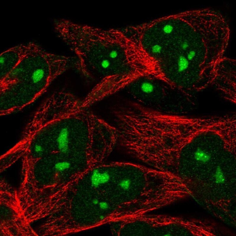
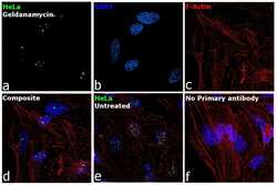
- Immunofluorescence analysis of WEE1 was performed using 70% confluent log phase HeLa treated with Geldanamycin (1 µM for 48 hours). The cells were fixed with 4% paraformaldehyde for 10 minutes, permeabilized with 0.1% Triton™ X-100 for 15 minutes, and blocked with 2% BSA for 45 minutes at room temperature. The cells were labeled with WEE1 Polyclonal Antibody (Product # PA5-67019) at 2 µg/mL dilution in 0.1% BSA, incubated at 4 degree celsius overnight and then labeled with Donkey anti-Rabbit IgG (H+L) Highly Cross-Adsorbed Secondary Antibody, Alexa Fluor Plus 488 (Product # A32790), (1:2000 dilution), for 45 minutes at room temperature (Panel a: Green). Nuclei (Panel b:Blue) were stained with ProLong™ Diamond Antifade Mountant with DAPI (Product # P36962). F-actin (Panel c: Red) was stained with Rhodamine Phalloidin (Product # R415, 1:300). Panel d represents the merged image showing predominantly nucleolar localization. Panel e represents untreated HeLa cells with higher expression levels for WEE1. Panel f represents control cells with no primary antibody to assess background. The images were captured at 60x magnification.

- Submitted by
- Invitrogen Antibodies (provider)
- Main image

- Experimental details
- Immunofluorescence analysis of WEE1 was performed using 70% confluent log phase HeLa treated with Geldanamycin (1 µM for 48 hours). The cells were fixed with 4% paraformaldehyde for 10 minutes, permeabilized with 0.1% Triton™ X-100 for 15 minutes, and blocked with 2% BSA for 45 minutes at room temperature. The cells were labeled with WEE1 Polyclonal Antibody (Product # PA5-67019) at 2 µg/mL dilution in 0.1% BSA, incubated at 4 degree Celsius overnight and then labeled with Donkey anti-Rabbit IgG (H+L) Highly Cross-Adsorbed Secondary Antibody, Alexa Fluor Plus 488 (Product # A32790), (1:2000 dilution), for 45 minutes at room temperature (Panel a: Green). Nuclei (Panel b:Blue) were stained with ProLong™ Diamond Antifade Mountant with DAPI (Product # P36962). F-actin (Panel c: Red) was stained with Rhodamine Phalloidin (Product # R415, 1:300). Panel d represents the merged image showing predominantly nucleolar localization. Panel e represents untreated HeLa cells with higher expression levels for WEE1. Panel f represents control cells with no primary antibody to assess background. The images were captured at 60x magnification.)