Explore
 Validate
 Learn
Learn Western blot
Antibody data
- Antibody Data
- Antigen structure
- References [0]
- Comments [0]
- Validations
- Western blot [7]
- Immunohistochemistry [1]
Submit
Validation data
Reference
Comment
Report error
- Product number
- PA5-29303 - Provider product page

- Provider
- Invitrogen Antibodies
- Product name
- WEE1 Polyclonal Antibody
- Antibody type
- Polyclonal
- Antigen
- Recombinant protein fragment
- Description
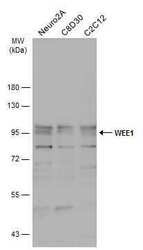
- Recommended positive controls: Jurkat, Raji, NCI-H929, Neuro2A, C8D30, C2C12. Predicted reactivity: Mouse (95%), Rat (96%), Xenopus laevis (84%), Chicken (88%), Bovine (98%). Store product as a concentrated solution. Centrifuge briefly prior to opening the vial.
- Reactivity
- Human, Mouse
- Host
- Rabbit
- Isotype
- IgG
- Vial size
- 100 µL
- Concentration
- 0.83 mg/mL
- Storage
- Store at 4°C short term. For long term storage, store at -20°C, avoiding freeze/thaw cycles.
No comments: Submit comment
Supportive validation
- Submitted by
- Invitrogen Antibodies (provider)
- Main image

- Experimental details
- Western blot analysis of WEE1 using 30 µg BCL-1 whole cell lysate. Samples were loaded onto a 7.5% SDS-PAGE gel and probed with a WEE1 polyclonal antibody (Product # PA5-29303) at a dilution of 1:1000.

- Submitted by
- Invitrogen Antibodies (provider)
- Main image
- Experimental details
- Western blot analysis of WEE1 using 30 µg of H1299 lysate. Samples were loaded onto a 7.5% SDS-PAGE gel and probed with a WEE1 polyclonal antibody (Product # PA5-29303) at a dilution of 1:1000.

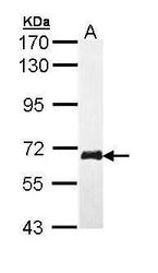
- Submitted by
- Invitrogen Antibodies (provider)
- Main image
- Experimental details
- Western Blot analysis of WEE1 was performed by separating 30 µg of various whole cell extracts by 7.5% SDS-PAGE. Proteins were transferred to a membrane and probed with a WEE1 Polyclonal Antibody (Product # PA5-29303) at a dilution of 1:500 and a HRP-conjugated anti-rabbit IgG secondary antibody.

- Submitted by
- Invitrogen Antibodies (provider)
- Main image

- Experimental details
- Western Blot using WEE1 Polyclonal Antibody (Product # PA5-29303). Various whole cell extracts (30 µg) were separated by 7.5% SDS-PAGE, and the membrane was blotted with WEE1 Polyclonal Antibody (Product # PA5-29303) diluted at 1:1,000. The HRP-conjugated anti-rabbit IgG antibody was used to detect the primary antibody.

- Submitted by
- Invitrogen Antibodies (provider)
- Main image

- Experimental details
- Knockdown of WEE1 was achieved by transfecting HeLa cells with WEE1 specific siRNA (Silencer® select Product # s21). Western blot analysis (Fig. a) was performed using whole cell extracts from the WEE1 knockdown cells (lane 3), non-specific scrambled siRNA transfected cells (lane 2) and untransfected cells (lane 1). The blot was probed with c-Cbl Polyclonal Antibody (Product # PA5-29303, 1:1000 dilution) and Goat anti-Rabbit IgG (H+L) Superclonal™ Recombinant Secondary Antibody, HRP (Product # A27036, 1:4000 dilution). Densitometric analysis of this western blot is shown in histogram (Fig. b). Decrease in signal upon siRNA mediated knock down confirms that antibody is specific to WEE1.

- Submitted by
- Invitrogen Antibodies (provider)
- Main image
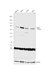
- Experimental details
- Western blot was performed using Anti-WEE1 polyclonal Antibody (Product # PA5-29303) and a 72 kDa band corresponding to WEE1 was observed in IMR-32, BeWo, Jurkat and HeLa. Whole cell extracts (30 µg lysate) of IMR-32 (Lane 1), BeWo (Lane 2), Jurkat (Lane 3) and HeLa (Lane 4) were electrophoresed using Novex® NuPAGE® 4-12 % Bis-Tris gel (Product # NP0322BOX). Resolved proteins were then transferred onto a nitrocellulose membrane (Product # IB23001) by iBlot® 2 Dry Blotting System (Product # IB21001). The blot was probed with the primary antibody (1:1000 dilution) and detected by chemiluminescence with Goat anti-Rabbit IgG (H+L) Superclonal™ Recombinant Secondary Antibody, HRP (Product # A27036, 1:4000 dilution) using the iBright FL 1000 (Product # A32752). Chemiluminescent detection was performed using Novex® ECL Chemiluminescent Substrate Reagent Kit (Product # WP20005).

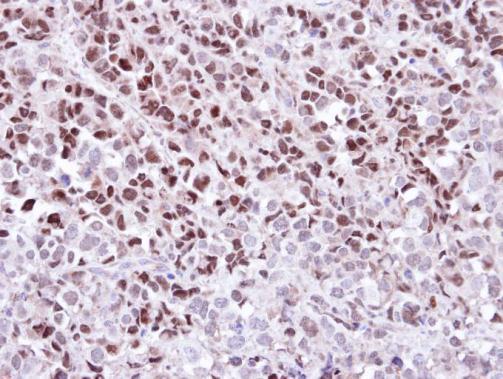
Supportive validation
- Submitted by
- Invitrogen Antibodies (provider)
- Main image
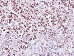
- Experimental details
- Immunohistochemical analysis of paraffin-embedded CL1-0 Xenograft, using WEE1 (Product # PA5-29303) antibody at 1:100 dilution. Antigen Retrieval: EDTA based buffer, pH 8.0, 15 min.