Explore
 Validate
 Learn
Learn Immunocytochemistry
Antibody data
- Antibody Data
- Antigen structure
- References [1]
- Comments [0]
- Validations
- Immunocytochemistry [1]
Submit
Validation data
Reference
Comment
Report error
- Product number
- HPA001056 - Provider product page

- Provider
- Atlas Antibodies
- Proper citation
- Atlas Antibodies Cat#HPA001056, RRID:AB_1845042
- Product name
- Anti-ARNT2
- Antibody type
- Polyclonal
- Description
- Polyclonal Antibody against Human ARNT2, Gene description: aryl-hydrocarbon receptor nuclear translocator 2, Alternative Gene Names: bHLHe1, KIAA0307, Validated applications: ICC, Uniprot ID: Q9HBZ2, Storage: Store at +4°C for short term storage. Long time storage is recommended at -20°C.
- Reactivity
- Human
- Host
- Rabbit
- Conjugate
- Unconjugated
- Isotype
- IgG
- Vial size
- 100 µl
- Concentration
- 0.1 mg/ml
- Storage
- Store at +4°C for short term storage. Long time storage is recommended at -20°C.
- Handling
- The antibody solution should be gently mixed before use.
Submitted references From Gene Expression Analysis to Tissue Microarrays
Ek S, Andréasson U, Hober S, Kampf C, Pontén F, Uhlén M, Merz H, Borrebaeck C
Molecular & Cellular Proteomics 2006;5(6):1072-1081
Molecular & Cellular Proteomics 2006;5(6):1072-1081
No comments: Submit comment
Supportive validation
- Submitted by
- Atlas Antibodies (provider)
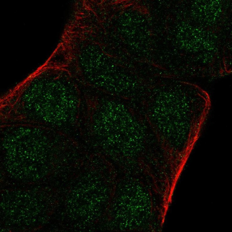
- Main image
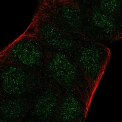
- Experimental details
- Immunofluorescent staining of human cell line RT4 shows localization to nucleus.
- Sample type
- Human