Explore
 Validate
 Learn
Learn Western blot
Antibody data
- Antibody Data
- Antigen structure
- References [0]
- Comments [0]
- Validations
- Western blot [1]
- Immunocytochemistry [1]
- Immunohistochemistry [1]
- Flow cytometry [1]
Submit
Validation data
Reference
Comment
Report error
- Product number
- F50622 - Provider product page

- Provider
- NSJ Bioreagents
- Product name
- Fgfr4 Antibody
- Antibody type
- Polyclonal
No comments: Submit comment
Supportive validation
- Submitted by
- NSJ Bioreagents (provider)
- Main image
- Experimental details
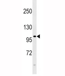
- FGFR4 antibody western blot analysis in 293 lysate. Observed molecular weight: 88~125 kDa depending on phosphorylation and glycosylation level.

Supportive validation
- Submitted by
- NSJ Bioreagents (provider)
- Main image
- Experimental details
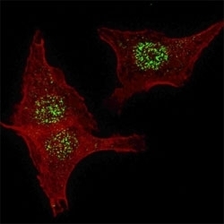
- Fluorescent confocal image of HeLa cells stained with FGFR4 antibody. HeLa cells were fixed with 4% PFA (20 min), permeabilized with Triton X-100 (0.2%, 30 min). Cells were then incubated with primary Ab (1:200, 2 h at room temperature). For secondary Ab, Alexa Fluor 488 conjugated donkey anti-rabbit Ab (green) was used (1:1000, 1h). Nuclei were counterstained with Hoechst 33342 (blue) (10 ug/ml, 5 min). Note the highly specific localization of FGFR4 to the nucleus, supported by Human Protein Atlas Data (http://www.proteinatlas.org/ENSG00000160867).

Supportive validation
- Submitted by
- NSJ Bioreagents (provider)
- Main image
- Experimental details
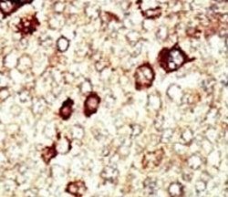
- IHC analysis of FFPE human hepatocarcinoma tissue stained with the FGFR4 antibody

Supportive validation
- Submitted by
- NSJ Bioreagents (provider)
- Main image
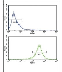
- Experimental details
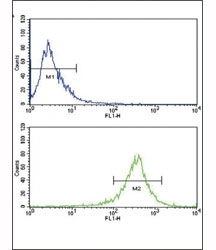
- Flow cytometric analysis of WiDr cells using FGFR4 antibody (bottom histogram) compared to a negative control (top histogram). FITC-conjugated goat-anti-rabbit secondary Ab was used for the analysis.