 Explore
 Validate
 Learn
Learn Immunohistochemistry
Antibody data
- Antibody Data
- Antigen structure
- References [7]
- Comments [0]
- Validations
- Immunohistochemistry [1]
- Flow cytometry [1]
- Other assay [1]
Submit
Validation data
Reference
Comment
Report error
- Product number
- 14-9768-80 - Provider product page

- Provider
- Invitrogen Antibodies
- Product name
- GRP78 Monoclonal Antibody (C38), eBioscience™
- Antibody type
- Monoclonal
- Antigen
- Other
- Description
- Description: The monoclonal antibody C38 recognizes human glucose-related protein 78 (GRP78). The GRP78 is encoded by the HSPA5 gene and functions as an endoplasmic reticulum (ER) chaperone protein and is a member of the heat shock protein 70 family. GRP78 functions within the ER lumen to facilitate proper protein folding, to prevent intermediates from aggregating, to target misfolded proteins for degradation, and as a calcium binding protein capable of recognizing ER stress and inducing the Unfolded Protein Response (UPR). During normal cellular function, GRP78 is bound to the transmembrane sensor proteins PERK, IRE1, and ATF6, keeping them in an inactive state. During ER stress, GRP78 binds to the hydrophobic residues of unfolded proteins within the ER and releases the sensor proteins, allowing signaling to the nucleus and the initiation of the UPR. During the UPR, protein synthesis is slowed and the transcription of ER chaperones is upregulated, increasing the capacity of the ER.
- Antibody clone number
- C38
- Concentration
- 0.5 mg/mL
Submitted references SDF4 Is a Prognostic Factor for 28-Days Mortality in Patients With Sepsis via Negatively Regulating ER Stress.
Cell surface GRP78 regulates BACE2 via lysosome-dependent manner to maintain mesenchymal phenotype of glioma stem cells.
Transplantation of iPS-Derived Tumor Cells with a Homozygous MHC Haplotype Induces GRP94 Antibody Production in MHC-Matched Macaques.
A murine monoclonal antibody directed against the carboxyl-terminal domain of GRP78 suppresses melanoma growth in mice.
Induction of unfolded protein response during neuronal induction of rat bone marrow stromal cells and mouse embryonic stem cells.
ER chaperones in mammalian development and human diseases.
GRP78 induction in cancer: therapeutic and prognostic implications.
Zhu T, Su Q, Wang C, Shen L, Chen H, Feng S, Peng X, Chen S, Wang Y, Jiang H, Chen J
Frontiers in immunology 2021;12:659193
Frontiers in immunology 2021;12:659193
Cell surface GRP78 regulates BACE2 via lysosome-dependent manner to maintain mesenchymal phenotype of glioma stem cells.
Chen Z, Wang H, Zhang Z, Xu J, Qi Y, Xue H, Gao Z, Zhao R, Wang S, Zhang S, Qiu W, Guo X, Li G
Journal of experimental & clinical cancer research : CR 2021 Jan 7;40(1):20
Journal of experimental & clinical cancer research : CR 2021 Jan 7;40(1):20
Transplantation of iPS-Derived Tumor Cells with a Homozygous MHC Haplotype Induces GRP94 Antibody Production in MHC-Matched Macaques.
Ishigaki H, Maeda T, Inoue H, Akagi T, Sasamura T, Ishida H, Inubushi T, Okahara J, Shiina T, Nakayama M, Itoh Y, Ogasawara K
Cancer research 2017 Nov 1;77(21):6001-6010
Cancer research 2017 Nov 1;77(21):6001-6010
A murine monoclonal antibody directed against the carboxyl-terminal domain of GRP78 suppresses melanoma growth in mice.
de Ridder GG, Ray R, Pizzo SV
Melanoma research 2012 Jun;22(3):225-35
Melanoma research 2012 Jun;22(3):225-35
Induction of unfolded protein response during neuronal induction of rat bone marrow stromal cells and mouse embryonic stem cells.
Cho YM, Jang YS, Jang YM, Chung SM, Kim HS, Lee JH, Jeong SW, Kim IK, Kim JJ, Kim KS, Kwon OJ
Experimental & molecular medicine 2009 Jun 30;41(6):440-52
Experimental & molecular medicine 2009 Jun 30;41(6):440-52
ER chaperones in mammalian development and human diseases.
Ni M, Lee AS
FEBS letters 2007 Jul 31;581(19):3641-51
FEBS letters 2007 Jul 31;581(19):3641-51
GRP78 induction in cancer: therapeutic and prognostic implications.
Lee AS
Cancer research 2007 Apr 15;67(8):3496-9
Cancer research 2007 Apr 15;67(8):3496-9
No comments: Submit comment
Supportive validation
- Submitted by
- Invitrogen Antibodies (provider)
- Main image
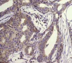
- Experimental details
- Immunohistochemistry of formalin-fixed paraffin embedded human breast cancer tissue using 5 µg/mL Anti-Human GRP78 Purified (right) followed by 10 µg/mL of Anti-Mouse IgG Biotin, Avidin HRP and DAB visualization.Nuclei are counterstained with hematoxylin.
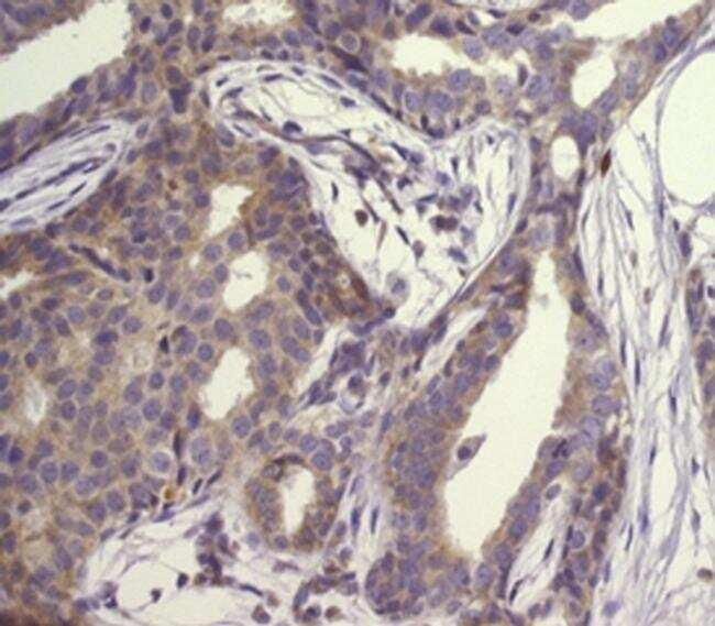
Supportive validation
- Submitted by
- Invitrogen Antibodies (provider)
- Main image
- Experimental details
- Intracellular staining of MCF7 with 1.0 µg Mouse IgG2b K Isotype Control Purified (Product # 14-4732-82) (blue) or 1.0 µg of Anti-Human GRP78 Purified (purple), followed by secondary staining with F (ab')2 Anti-Mouse IgG PE (Product # 12-4010-82).Cells were fixed and permeabilized using the Intracellular Fixation & Permeabilization Buffer Set (Product # 88-8824-00).Viable cells were used for analysis.

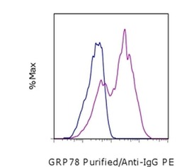
Supportive validation
- Submitted by
- Invitrogen Antibodies (provider)
- Main image
- Experimental details
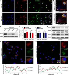
- Fig. 6 Blocking csGRP78 induces lysosomal degradation of BACE2. a Co-immunofluorescence staining of csGRP78 (red) and BACE2 (green) in GSC20 and 267. Scale bar, 25 mum. b Colocalization analysis for co-expression of BACE2 and csGRP78 in cell membrane of GSC20 and 267 using colocalization finder plugin. c Western blotting for BACE2 in GSC267 with anti-GRP78 treatment for 72 h or lentiviral shGRP78 expressing. d BACE2 RNA level in GSC20 and 267 was detected by qRT-PCR with anti-GRP78 treatment or lentiviral shGRP78 expressing, GADPH as the reference gene. e Western blotting for BACE2 protein in GSC267 that treated with MG-132, Chloroquine (CQ) or anti-GRP78, respectively, and co-treatment of anti-GRP78 with MG-132 or CQ. f and g Upper, the confocal microscopy images for overview and splitting channel of BACE2 (red) and LAMP1 (green). The yellow triangles indicate the overlapped regions (yellow). Lower, the analysis plot for the fluorescence intensity of two channels. Red line for BACE2 and green line for LAMP1. Scale bar, 5 mum, and 25 mum for the large field of view. Error bar indicates at least three independent experiments and data are shown as mean +- SD
