 Explore
 Validate
 Learn
Learn Immunocytochemistry
Antibody data
- Antibody Data
- Antigen structure
- References [2]
- Comments [0]
- Validations
- Immunocytochemistry [1]
- Other assay [2]
Submit
Validation data
Reference
Comment
Report error
- Product number
- PA1-014A-A488 - Provider product page

- Provider
- Invitrogen Antibodies
- Product name
- GRP78 Polyclonal Antibody, Alexa Fluor™ 488
- Antibody type
- Polyclonal
- Antigen
- Synthetic peptide
- Description
- PA1014AA488 detects glucose regulated protein/BiP (GRP78) human, rat and mouse samples. The PA1014AA488 immunogen is a synthetic peptide corresponding to residues C T(643) G E E D T S E K D E L(654) of rat GRP78.
- Reactivity
- Human, Mouse, Rat
- Host
- Rabbit
- Conjugate
- Green dye
- Isotype
- IgG
- Vial size
- 50 µL
- Concentration
- 1 mg/mL
- Storage
- 4° C, store in dark, DO NOT FREEZE!
Submitted references Cytokine-induced translocation of GRP78 to the plasma membrane triggers a pro-apoptotic feedback loop in pancreatic beta cells.
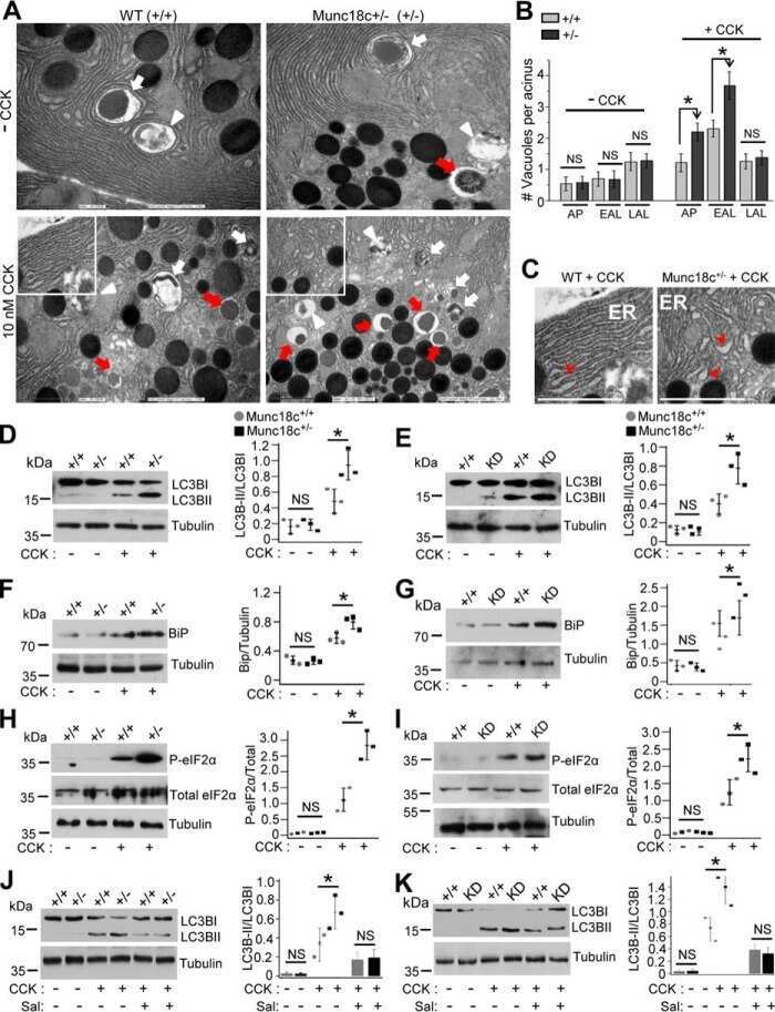
Depletion of the membrane-fusion regulator Munc18c attenuates caerulein hyperstimulation-induced pancreatitis.
Vig S, Buitinga M, Rondas D, Crèvecoeur I, van Zandvoort M, Waelkens E, Eizirik DL, Gysemans C, Baatsen P, Mathieu C, Overbergh L
Cell death & disease 2019 Apr 5;10(4):309
Cell death & disease 2019 Apr 5;10(4):309
Depletion of the membrane-fusion regulator Munc18c attenuates caerulein hyperstimulation-induced pancreatitis.
Dolai S, Liang T, Orabi AI, Xie L, Holmyard D, Javed TA, Fernandez NA, Xie H, Cattral MS, Thurmond DC, Thorn P, Gaisano HY
The Journal of biological chemistry 2018 Feb 16;293(7):2510-2522
The Journal of biological chemistry 2018 Feb 16;293(7):2510-2522
No comments: Submit comment
Supportive validation
- Submitted by
- Invitrogen Antibodies (provider)
- Main image
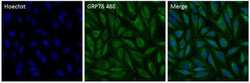
- Experimental details
- Immunofluorescent analysis of Grp78 (green) in HeLa cells. The cells were fixed with 4% Paraformaldehyde in PBS for 15 minutes at room temperature, and blocked with 3% BSA in PBS (Product # 37525) for 30 minutes at room temperature. Cells were stained with a Grp78 Polyclonal Antibody, AlexaFluor 488 conjugate (Product # PA1-014A-A488) at a dilution of 2.5 µg/mL in blocking buffer for 1 hour at room temperature protected from light. Nuclei (blue) were stained with Hoechst Dye (Product # 62249) at a dilution of 1:10,000 in blocking buffer. Images were taken on a Thermo Scientific ToxInsight Instrument at 20X magnification.
- Conjugate
- Green dye

Supportive validation
- Submitted by
- Invitrogen Antibodies (provider)
- Main image
- Experimental details
- NULL
- Conjugate
- Green dye
- Submitted by
- Invitrogen Antibodies (provider)
- Main image

- Experimental details
- Fig. 2 Surface translocation of glucose-regulated protein 78 (GRP78) in beta cells is an early phenomenon, taking place in the early apoptotic cell population. MIN6 cells exposed to cytokines (human interleukin-1beta (50 U/mL), mouse interferon-gamma (250 U/mL), and mouse tumor necrosis factor-alpha (1000 U/mL)) for the indicated time points (4, 8, 16, and 24 h) were stained with the late-apoptosis/necrosis marker DRAQ7-AF700, early apoptosis marker Annexin V-APC (see Results for the interpretation of these combined dyes to discriminate between living, early, and late apoptotic cells), and anti-GRP78-FITC, followed by flow cytometry. a , b Percentage of living ( a ) and early apoptotic ( b ) cell population in control (C) and cytokine (Cyt) exposed MIN6 cells. c , d Percentage of sGRP78-positive cells in the living ( c ) and early apoptotic ( d ) subpopulation. e , f Geometric mean fluorescence intensity (MFI) for sGRP78 in the living ( e ) and early apoptotic ( f ) subpopulation. Data are presented as mean +- SEM ( n = 4) and statistically analyzed by a two-way analysis of variance followed by Sidak posthoc test for multiple group comparisons. * P < 0.05, ** P < 0.01, *** P < 0.001, and **** P < 0.0001
- Conjugate
- Green dye
