 Explore
 Validate
 Learn
Learn Western blot
 Immunohistochemistry
Antibody data
- Antibody Data
- Antigen structure
- References [2]
- Comments [0]
- Validations
- Western blot [5]
- Flow cytometry [4]
- Other assay [1]
Submit
Validation data
Reference
Comment
Report error
- Product number
- PA5-22967 - Provider product page

- Provider
- Invitrogen Antibodies
- Product name
- GRP78 Polyclonal Antibody
- Antibody type
- Polyclonal
- Antigen
- Synthetic peptide
- Description
- This antibody is predicted to react with zebrafish, c. elegans, primate, bovine, and drosophila based on 100% sequence homology. Suggested positive control: HeLa whole cell extract.
- Reactivity
- Human, Mouse, Rat, Chicken/Avian, Drosophila, Zebrafish
- Host
- Rabbit
- Isotype
- IgG
- Vial size
- 100 µL
- Concentration
- 1.0 mg/mL
- Storage
- Store at 4°C short term. For long term storage, store at -20°C, avoiding freeze/thaw cycles.
Submitted references Gibberellic Acid Initiates ER Stress and Activation of Differentiation in Cultured Human Immortalized Keratinocytes HaCaT and Epidermoid Carcinoma Cells A431.
Conserved pharmacological rescue of hereditary spastic paraplegia-related phenotypes across model organisms.
Vildanova M, Vishnyakova P, Saidova A, Konduktorova V, Onishchenko G, Smirnova E
Pharmaceutics 2021 Oct 30;13(11)
Pharmaceutics 2021 Oct 30;13(11)
Conserved pharmacological rescue of hereditary spastic paraplegia-related phenotypes across model organisms.
Julien C, Lissouba A, Madabattula S, Fardghassemi Y, Rosenfelt C, Androschuk A, Strautman J, Wong C, Bysice A, O'sullivan J, Rouleau GA, Drapeau P, Parker JA, Bolduc FV
Human molecular genetics 2016 Mar 15;25(6):1088-99
Human molecular genetics 2016 Mar 15;25(6):1088-99
No comments: Submit comment
Supportive validation
- Submitted by
- Invitrogen Antibodies (provider)
- Main image

- Experimental details
- Western blot analysis of GRP78 using a polyclonal antibody (Product # PA5-22967).

- Submitted by
- Invitrogen Antibodies (provider)
- Main image

- Experimental details
- Western blot analysis was performed on whole cell extract (30 µg lysate) of MCF7 (Lane 1), MCF7 treated with Thapsigargin (1uM, 24 hrs) (Lane 2), HeLa (Lane 3), HeLa treated with Thapsigargin (1uM, 24 hrs) (Lane 4), MOLT-4 (Lane 5), and MOLT-4 treated with Thapsigargin (1uM, 24 hrs) (Lane 6). The blot was probed with Anti-GRP78 Polyclonal Antibody (Product # PA5-22967, 1:2000 dilution) and detected by chemiluminescence using Goat anti-Rabbit IgG (H+L) Superclonal™ Secondary Antibody, HRP conjugate (Product # A27036, 0.25 µg/ml, 1:4000 dilution). A 78 kDa band corresponding to GRP78 was observed in all cell lines tested and was enhanced upon Thapsigargin treatment.

- Submitted by
- Invitrogen Antibodies (provider)
- Main image

- Experimental details
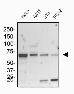
- Western blot analysis of GRP78 in human HeLa and A431 cells, mouse 3T3 cells and rat PC12 cells. Samples were incubated in GRP78 polyclonal antibody (Product # PA5-22967) using a dilution of 1.0 µg/mL followed by an anti-rabbit HRP secondary antibody. separated on a 7.5% gel by SDS-PAGE, transferred to PVDF membrane and blocked in 5% non-fat milk in TBST. Detection: chemiluminescence.
- Submitted by
- Invitrogen Antibodies (provider)
- Main image

- Experimental details
- Western blot analysis of GRP78 in HeLa whole cell extracts. Sample was incubated in GRP78 polyclonal antibody (Product # PA5-22967).

- Submitted by
- Invitrogen Antibodies (provider)
- Main image

- Experimental details
- Western blot analysis of GRP78 in 0.1 mg/mL HeLa lysate. Samples were incubated in GRP78 polyclonal antibody (Product # PA5-22967). This experiment was performed under reducing conditions using the 12-230 kDa separation system.

Supportive validation
- Submitted by
- Invitrogen Antibodies (provider)
- Main image

- Experimental details
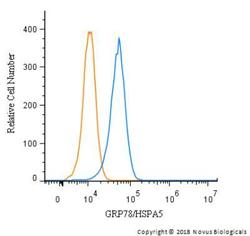
- Flow cytometry of GRP78 in HeLa cells (blue) and a matched isotype control (orange). Samples were incubated in GRP78 polyclonal antibody (Product # PA5-22967) using a dilution of 1 µg of antibody added to 100 µL of staining buffer and cells were incubated for 30 minutes at room temperature. Cells were fixed with 4% PFA and then permeabilized with 0.1% saponin. Both antibodies were conjugated to Alexa Fluor 488.

- Submitted by
- Invitrogen Antibodies (provider)
- Main image

- Experimental details
- Flow cytometry of GRP78 in Jurkat cells. Samples were incubated in GRP78 polyclonal (Product # PA5-22967) using a dilution of 2 µg/mL for 30 minutes at room temperature. Antibody (blue) and a matched isotype control (orange). Cells were fixed with 4% PFA and then permeablized with 0.1% saponin. Both antibodies were conjugated to Alexa Fluor 647.

- Submitted by
- Invitrogen Antibodies (provider)
- Main image

- Experimental details
- Flow cytometry of GRP78 in NIH3T3 cells (blue) and a matched isotype control (orange). Samples were incubated in GRP78 polyclonal antibody (Product # PA5-22967) using a dilution of 5 µg/mL for 30 minutes at room temperature. Cells were fixed with 4% PFA and then permeabilized with 0.1% saponin. Both antibodies were conjugated to Alexa Fluor 488.

- Submitted by
- Invitrogen Antibodies (provider)
- Main image
- Experimental details
- Flow cytometry of GRP78 in HeLa with and a matched isotype control. Samples were incubated in GRP78 polyclonal antibody (Product # PA5-22967) using a dilution of 1 µg/mL for 30 minutes at room temperature followed by a Rabbit IgG (H+L) Cross-Adsorbed Secondary Antibody, Dylight™ 550. Cells were fixed with 4% PFA and then permeabilized with 0.1% saponin.

Supportive validation
- Submitted by
- Invitrogen Antibodies (provider)
- Main image

- Experimental details
- Figure 2 Immunocytochemical visualization of GRP78 localization and analysis of GRP78 content in keratinocytes HaCaT and carcinoma A431 cells. ( a - i ) Keratinocytes HaCaT in control specimens ( a - c ) and in the presence of DTT ( d - f ) and GA ( g , h ); ( j - r ) carcinoma A431 cells in control specimens ( j - l ), in the presence of DTT ( m - o ) and GA ( p - r ). Nuclei are stained with DAPI (left column); GRP78 is visualized with anti-GRP78 antibodies (middle column), merged images (right column). Scale bar, 20 mum. Western blot analysis and evaluation of GRP78 content in HaCaT ( s ) and A431 cells ( t ). C1--cells growing with ethanol as a solvent for GA (control for GA); C2--cells growing in standard conditions (control for DTT). Data are shown as mean +- standard deviation ( n = 3-5); * p
