Explore
 Validate
 Learn
Learn Western blot
Antibody data
- Antibody Data
- Antigen structure
- References [1]
- Comments [0]
- Validations
- Western blot [4]
- Immunocytochemistry [1]
- Immunohistochemistry [3]
Submit
Validation data
Reference
Comment
Report error
- Product number
- GTX127934 - Provider product page

- Provider
- GeneTex
- Product name
- Grp78 antibody
- Antibody type
- Polyclonal
- Reactivity
- Human, Mouse, Rat
- Host
- Rabbit
Submitted references Extracellular vesicles with altered tetraspanin CD9 and CD151 levels confer increased prostate cell motility and invasion.
Brzozowski JS, Bond DR, Jankowski H, Goldie BJ, Burchell R, Naudin C, Smith ND, Scarlett CJ, Larsen MR, Dun MD, Skelding KA, Weidenhofer J
Scientific reports 2018 Jun 11;8(1):8822
Scientific reports 2018 Jun 11;8(1):8822
No comments: Submit comment
Supportive validation
- Submitted by
- GeneTex (provider)
- Main image
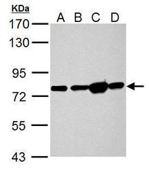
- Experimental details
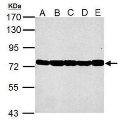
- Hspa5 antibody detects Hspa5 protein by Western blot analysis.A. 50 ?g Rat heart lysate/extractB. 50 ?g Rat Lung lysate/extractC. 50 ?g Rat Liver lysate/extractD. 50 ?g Rat kidney lysate/extract7.5 % SDS-PAGEHspa5 antibody (GTX127934) dilution: 1:10000
- Submitted by
- GeneTex (provider)
- Main image
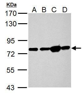
- Experimental details
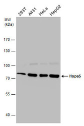
- Sample (30 ug of whole cell lysate) A: 293T B: A431 C: HeLa D: HepG2 E: A375 7.5% SDS PAGE GTX127934 diluted at 1:10000

- Submitted by
- GeneTex (provider)
- Main image
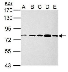
- Experimental details
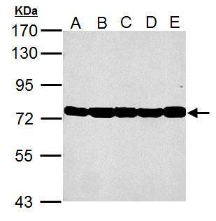
- Sample (30 ug of whole cell lysate) A: NIH-3T-3 B: JC C: BCL-1 D: C2C12 E: Raw264.7 7.5% SDS PAGE GTX127934 diluted at 1:10000
- Submitted by
- GeneTex (provider)
- Main image
- Experimental details
- Hspa5 antibody detects Hspa5 protein by Western blot analysis. Various whole cell extracts (30 £gg) were separated by 7.5% SDS-PAGE, and the membrane was blotted with Hspa5 antibody (GTX127934) diluted at a dilution of 1:10000.

Supportive validation
- Submitted by
- GeneTex (provider)
- Main image

- Experimental details
- Hspa5 antibody detects Hspa5 protein at endoplasmic reticulum by immunofluorescent analysis.Sample: HeLa cells were fixed in ice-cold MeOH for 5 min.Green: Hspa5 protein stained by Hspa5 antibody (GTX127934) diluted at 1:500.Blue: Hoechst 33342 staining.Scale bar = 10 £gm.

Supportive validation
- Submitted by
- GeneTex (provider)
- Main image
- Experimental details
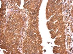
- Hspa5 antibody detects Hspa5 protein at cytosol on human gastric cancer by immunohistochemical analysis. Sample: Paraffin-embedded gastric cancer. Hspa5 antibody (GTX127934) dilution: 1:500.

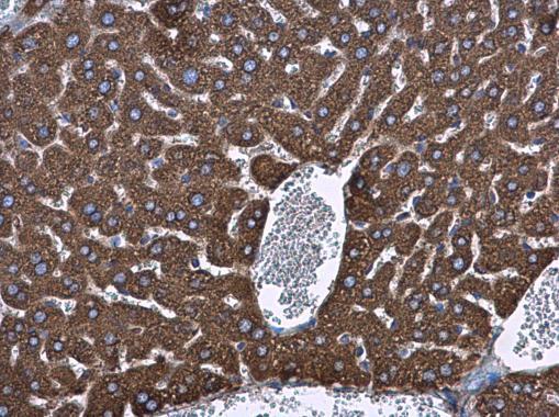
- Submitted by
- GeneTex (provider)
- Main image
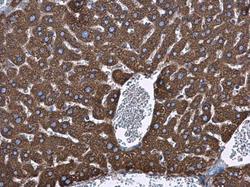
- Experimental details
- Hspa5 antibody detects Hspa5 protein at cytoplasm in mouse liver by immunohistochemical analysis. Sample: Paraffin-embedded mouse liver. Hspa5 antibody (GTX127934) diluted at 1:500.
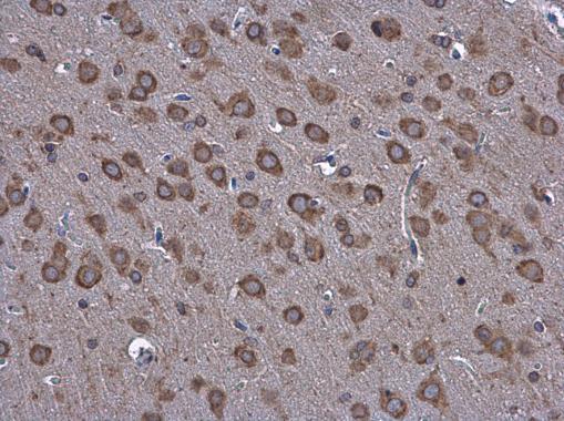
- Submitted by
- GeneTex (provider)
- Main image
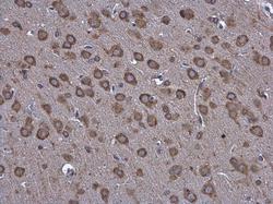
- Experimental details
- Hspa5 antibody detects Hspa5 protein at cytoplasm in rat brain by immunohistochemical analysis. Sample: Paraffin-embedded rat brain. Hspa5 antibody (GTX127934) diluted at 1:500.