Explore
 Validate
 Learn
Learn Western blot
Antibody data
- Antibody Data
- Antigen structure
- References [5]
- Comments [0]
- Validations
- Western blot [2]
Submit
Validation data
Reference
Comment
Report error
- Product number
- AF4846 - Provider product page

- Provider
- R&D Systems
- Product name
- Human/Mouse/Rat GRP78/HSPA5 Antibody
- Antibody type
- Polyclonal
- Description
- Immunogen affinity purified. Detects endogenous human, mouse and rat GRP78 in Western blots.
- Reactivity
- Human, Mouse, Rat
- Host
- Goat
- Conjugate
- Unconjugated
- Antigen sequence
P11021- Isotype
- IgG
- Vial size
- 100 ug
- Concentration
- LYOPH
- Storage
- Use a manual defrost freezer and avoid repeated freeze-thaw cycles. 12 months from date of receipt, -20 to -70 °C as supplied. 1 month, 2 to 8 °C under sterile conditions after reconstitution. 6 months, -20 to -70 °C under sterile conditions after reconstitution.
Submitted references XBP1 signalling is essential for alleviating mutant protein aggregation in ER-stress related skeletal disease.
The Role of Autophagy in the Degradation of Misfolded HLA-B27 Heavy Chains.
The Novel Mechanisms Concerning the Inhibitions of Palmitate-Induced Proinflammatory Factor Releases and Endogenous Cellular Stress with Astaxanthin on MIN6 β-Cells.
Open reading frame 3 of genotype 1 hepatitis E virus inhibits nuclear factor-κappa B signaling induced by tumor necrosis factor-α in human A549 lung epithelial cells.
Overexpression of TGF-ß 1 gene induces cell surface localized glucose-regulated protein 78-associated latency-associated peptide/TGF-ß.
Piróg KA, Dennis EP, Hartley CL, Jackson RM, Soul J, Schwartz JM, Bateman JF, Boot-Handford RP, Briggs MD
PLoS genetics 2019 Jul;15(7):e1008215
PLoS genetics 2019 Jul;15(7):e1008215
The Role of Autophagy in the Degradation of Misfolded HLA-B27 Heavy Chains.
Navid F, Layh-Schmitt G, Sikora KA, Cougnoux A, Colbert RA
Arthritis & rheumatology (Hoboken, N.J.) 2018 May;70(5):746-755
Arthritis & rheumatology (Hoboken, N.J.) 2018 May;70(5):746-755
The Novel Mechanisms Concerning the Inhibitions of Palmitate-Induced Proinflammatory Factor Releases and Endogenous Cellular Stress with Astaxanthin on MIN6 β-Cells.
Kitahara A, Takahashi K, Morita N, Murashima T, Onuma H, Sumitani Y, Tanaka T, Kondo T, Hosaka T, Ishida H
Marine drugs 2017 Jun 20;15(6)
Marine drugs 2017 Jun 20;15(6)
Open reading frame 3 of genotype 1 hepatitis E virus inhibits nuclear factor-κappa B signaling induced by tumor necrosis factor-α in human A549 lung epithelial cells.
Xu J, Wu F, Tian D, Wang J, Zheng Z, Xia N
PloS one 2014;9(6):e100787
PloS one 2014;9(6):e100787
Overexpression of TGF-ß 1 gene induces cell surface localized glucose-regulated protein 78-associated latency-associated peptide/TGF-ß.
Oida T, Weiner HL
Journal of immunology (Baltimore, Md. : 1950) 2010 Sep 15;185(6):3529-35
Journal of immunology (Baltimore, Md. : 1950) 2010 Sep 15;185(6):3529-35
No comments: Submit comment
Supportive validation
- Submitted by
- R&D Systems (provider)
- Main image
- Experimental details
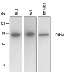
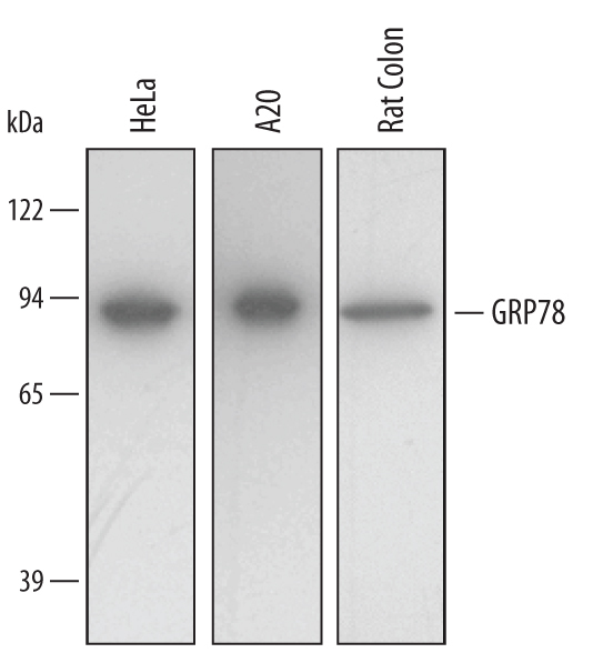
- Detection of Human and Rat GRP78/HSPA5 by Western Blot. Western blot shows lysates of HeLa human cervical epithelial carcinoma cell line and A20 mouse B cell lymphoma cell line. PVDF membrane was probed with 0.5 µg/mL of Human/Mouse/Rat GRP78/HSPA5 Antigen Affinity-purified Polyclonal Antibody (Catalog # AF4846) followed by HRP-conjugated Anti-Goat IgG Secondary Antibody (Catalog # HAF109). A specific band was detected for GRP78/HSPA5 at approximately 80 kDa (as indicated). This experiment was conducted under reducing conditions and using Immunoblot Buffer Group 2.
- Submitted by
- R&D Systems (provider)
- Main image

- Experimental details
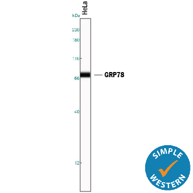
- Detection of Human GRP78/HSPA5 by Simple WesternTM. Simple Western lane view shows lysates of HeLa human cervical epithelial carcinoma cell line, loaded at 0.2 mg/mL. A specific band was detected for GRP78/HSPA5 at approximately 74 kDa (as indicated) using 5 µg/mL of Goat Anti-Human/Mouse/Rat GRP78/HSPA5 Antigen Affinity-purified Polyclonal Antibody (Catalog # AF4846) followed by 1:50 dilution of HRP-conjugated Anti-Goat IgG Secondary Antibody (Catalog # HAF109). This experiment was conducted under reducing conditions and using the 12-230 kDa separation system.