 Explore
 Validate
 Learn
Learn Western blot
Antibody data
- Antibody Data
- Antigen structure
- References [3]
- Comments [0]
- Validations
- Western blot [2]
Submit
Validation data
Reference
Comment
Report error
- Product number
- MAB4846 - Provider product page

- Provider
- R&D Systems
- Product name
- Human GRP78/HSPA5 Antibody
- Antibody type
- Monoclonal
- Description
- Protein A or G purified from hybridoma culture supernatant. Detects endogenous human GRP78 at 78 kDa in Western blots.
- Reactivity
- Human
- Host
- Mouse
- Conjugate
- Unconjugated
- Antigen sequence
P11021- Isotype
- IgG
- Antibody clone number
- 474421
- Vial size
- 100 ug
- Concentration
- LYOPH
- Storage
- Use a manual defrost freezer and avoid repeated freeze-thaw cycles. 12 months from date of receipt, -20 to -70 °C as supplied. 1 month, 2 to 8 °C under sterile conditions after reconstitution. 6 months, -20 to -70 °C under sterile conditions after reconstitution.
Submitted references Middle East respiratory syndrome coronavirus and bat coronavirus HKU9 both can utilize GRP78 for attachment onto host cells.
WWOX sensitises ovarian cancer cells to paclitaxel via modulation of the ER stress response.
The endoplasmic reticulum chaperone BiP/GRP78 is important in the structure and function of the human cytomegalovirus assembly compartment.
Chu H, Chan CM, Zhang X, Wang Y, Yuan S, Zhou J, Au-Yeung RK, Sze KH, Yang D, Shuai H, Hou Y, Li C, Zhao X, Poon VK, Leung SP, Yeung ML, Yan J, Lu G, Jin DY, Gao GF, Chan JF, Yuen KY
The Journal of biological chemistry 2018 Jul 27;293(30):11709-11726
The Journal of biological chemistry 2018 Jul 27;293(30):11709-11726
WWOX sensitises ovarian cancer cells to paclitaxel via modulation of the ER stress response.
Janczar S, Nautiyal J, Xiao Y, Curry E, Sun M, Zanini E, Paige AJ, Gabra H
Cell death & disease 2017 Jul 27;8(7):e2955
Cell death & disease 2017 Jul 27;8(7):e2955
The endoplasmic reticulum chaperone BiP/GRP78 is important in the structure and function of the human cytomegalovirus assembly compartment.
Buchkovich NJ, Maguire TG, Paton AW, Paton JC, Alwine JC
Journal of virology 2009 Nov;83(22):11421-8
Journal of virology 2009 Nov;83(22):11421-8
No comments: Submit comment
Supportive validation
- Submitted by
- R&D Systems (provider)
- Main image
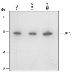
- Experimental details
- Detection of Human GRP78/HSPA5 by Western Blot. Western blot shows lysates of HeLa human cervical epithelial carcinoma cell line, Jurkat human acute T cell leukemia cell line, and MCF-7 human breast cancer cell line. PVDF membrane was probed with 1 µg/mL of Human GRP78/HSPA5 Monoclonal Antibody (Catalog # MAB4846) followed by HRP-conjugated Anti-Mouse IgG Secondary Antibody (Catalog # HAF007). A specific band was detected for GRP78/HSPA5 at approximately 78 kDa (as indicated). This experiment was conducted under reducing conditions and using Immunoblot Buffer Group 2.
- Submitted by
- R&D Systems (provider)
- Main image
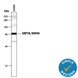
- Experimental details
- Detection of Human GRP78/HSPA5 by Simple WesternTM. Simple Western lane view shows lysates of Jurkat human acute T cell leukemia cell line, loaded at 0.2 mg/mL. A specific band was detected for GRP78/HSPA5 at approximately 72 kDa (as indicated) using 10 µg/mL of Mouse Anti-Human GRP78/HSPA5 Monoclonal Antibody (Catalog # MAB4846). This experiment was conducted under reducing conditions and using the 12-230 kDa separation system. *Non-specific interaction with the 230 kDa Simple Western standard may be seen with this antibody.
