Explore
 Validate
 Learn
Learn Western blot
Antibody data
- Antibody Data
- Antigen structure
- References [4]
- Comments [0]
- Validations
- Western blot [1]
- Immunohistochemistry [1]
Submit
Validation data
Reference
Comment
Report error
- Product number
- sc-9090 - Provider product page

- Provider
- Santa Cruz Biotechnology
- Proper citation
- Santa Cruz Biotechnology Cat#sc-9090, RRID:AB_2129021
- Product name
- Anti-ITGB4
- Antibody type
- Polyclonal
- Antigen
- Recombinant full-length protein
- Reactivity
- Human
- Host
- Rabbit
Submitted references Tumor cell invasiveness correlates with changes in integrin expression and localization
Role of binding of plectin to the integrin beta4 subunit in the assembly of hemidesmosomes.
Beta4 integrin and laminin 5 are aberrantly expressed in polycystic kidney disease: role in increased cell adhesion and migration.
Ras and TGF[beta] cooperatively regulate epithelial cell plasticity and metastasis: dissection of Ras signaling pathways.
Sabine Maschler, Gerhard Wirl, Herbert Spring, Dorothea v Bredow, Isabelle Sordat, Hartmut Beug, Ernst Reichmann
Oncogene 2005 Jan;24(12):2032-2041
Oncogene 2005 Jan;24(12):2032-2041
Role of binding of plectin to the integrin beta4 subunit in the assembly of hemidesmosomes.
Koster J, van Wilpe S, Kuikman I, Litjens SH, Sonnenberg A
Molecular biology of the cell 2004 Mar;15(3):1211-23
Molecular biology of the cell 2004 Mar;15(3):1211-23
Beta4 integrin and laminin 5 are aberrantly expressed in polycystic kidney disease: role in increased cell adhesion and migration.
Joly D, Morel V, Hummel A, Ruello A, Nusbaum P, Patey N, Noël LH, Rousselle P, Knebelmann B
The American journal of pathology 2003 Nov;163(5):1791-800
The American journal of pathology 2003 Nov;163(5):1791-800
Ras and TGF[beta] cooperatively regulate epithelial cell plasticity and metastasis: dissection of Ras signaling pathways.
Janda E, Lehmann K, Killisch I, Jechlinger M, Herzig M, Downward J, Beug H, Grünert S
The Journal of cell biology 2002 Jan 21;156(2):299-313
The Journal of cell biology 2002 Jan 21;156(2):299-313
No comments: Submit comment
Supportive validation
- Submitted by
- per
- Main image
- Experimental details
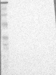
- Western blot analysis of antibody specificity using a routine panel composed of IgG/HSA-depleted human plasma and protein lysates from selected human tissues and cell lines.
- Validation comment
- No bands detected.
- Primary Ab dilution
- 1:500
- Secondary Ab dilution
- 1:3000
- Lane 1
- Marker [kDa]: 229, 112, 83.5, 47.9, 32.3, 26.5, 17.2
- Lane 2
- RT-4
- Lane 3
- U-251MG sp
- Lane 4
- Human Plasma
- Lane 5
- Liver
- Lane 6
- Tonsil
- Theoretical target weight
- [kDa] 202

Supportive validation
- Submitted by
- per
- Main image
- Experimental details
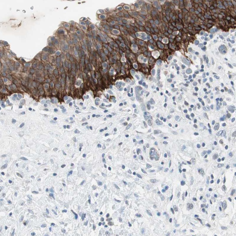
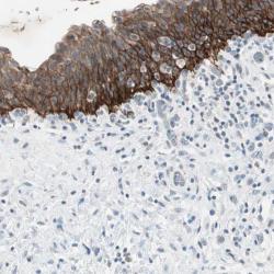
- Immunohistochemical staining of human urinary bladder shows distinct membranous and cytoplasmic positivity in urothelial cells.
- Validation comment
- Staining pattern consistent with experimental and/or bioinformatic data.