Explore
 Validate
 Learn
Learn Western blot
 Immunocytochemistry
Antibody data
- Antibody Data
- Antigen structure
- References [3]
- Comments [0]
- Validations
- Immunocytochemistry [1]
Submit
Validation data
Reference
Comment
Report error
- Product number
- HPA036348 - Provider product page

- Provider
- Atlas Antibodies
- Proper citation
- Atlas Antibodies Cat#HPA036348, RRID:AB_2675077
- Product name
- Anti-ITGB4
- Antibody type
- Polyclonal
- Description
- Polyclonal Antibody against Human ITGB4, Gene description: integrin, beta 4, Alternative Gene Names: CD104, Validated applications: ICC, IHC, WB, Uniprot ID: P16144, Storage: Store at +4°C for short term storage. Long time storage is recommended at -20°C.
- Reactivity
- Human
- Host
- Rabbit
- Conjugate
- Unconjugated
- Isotype
- IgG
- Vial size
- 100 µl
- Concentration
- 0.1 mg/ml
- Storage
- Store at +4°C for short term storage. Long time storage is recommended at -20°C.
- Handling
- The antibody solution should be gently mixed before use.
Submitted references Chaperone-Mediated Autophagy Controls Proteomic and Transcriptomic Pathways to Maintain Glioma Stem Cell Activity
Identification of integrin drug targets for 17 solid tumor types
Analysis of risk factors for post-operative complications and prognostic predictors of disease recurrence following definitive treatment of patients with esophageal cancer from two medical centers in Northwest China
Auzmendi-Iriarte J, Otaegi-Ugartemendia M, Carrasco-Garcia E, Azkargorta M, Diaz A, Saenz-Antoñanzas A, Andermatten J, Garcia-Puga M, Garcia I, Elua-Pinin A, Ruiz I, Sampron N, Elortza F, Cuervo A, Matheu A
Cancer Research 2022;82(7):1283-1297
Cancer Research 2022;82(7):1283-1297
Identification of integrin drug targets for 17 solid tumor types
Arun A, Tepper C, Lam K
Oncotarget 2018;9(53):30146-30162
Oncotarget 2018;9(53):30146-30162
Analysis of risk factors for post-operative complications and prognostic predictors of disease recurrence following definitive treatment of patients with esophageal cancer from two medical centers in Northwest China
Wang J, Zhang B, Meng J, Xiao G, Li X, Li G, Qin S, Du N, Zhang J, Zhang J, Xu C, Tang S, Liang R, Ren H, Sun X
Experimental and Therapeutic Medicine 2017;14(3):2584-2594
Experimental and Therapeutic Medicine 2017;14(3):2584-2594
No comments: Submit comment
Supportive validation
- Submitted by
- Atlas Antibodies (provider)
- Main image
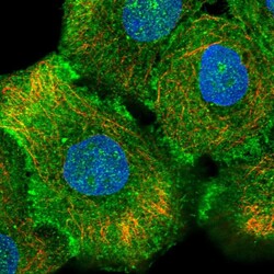
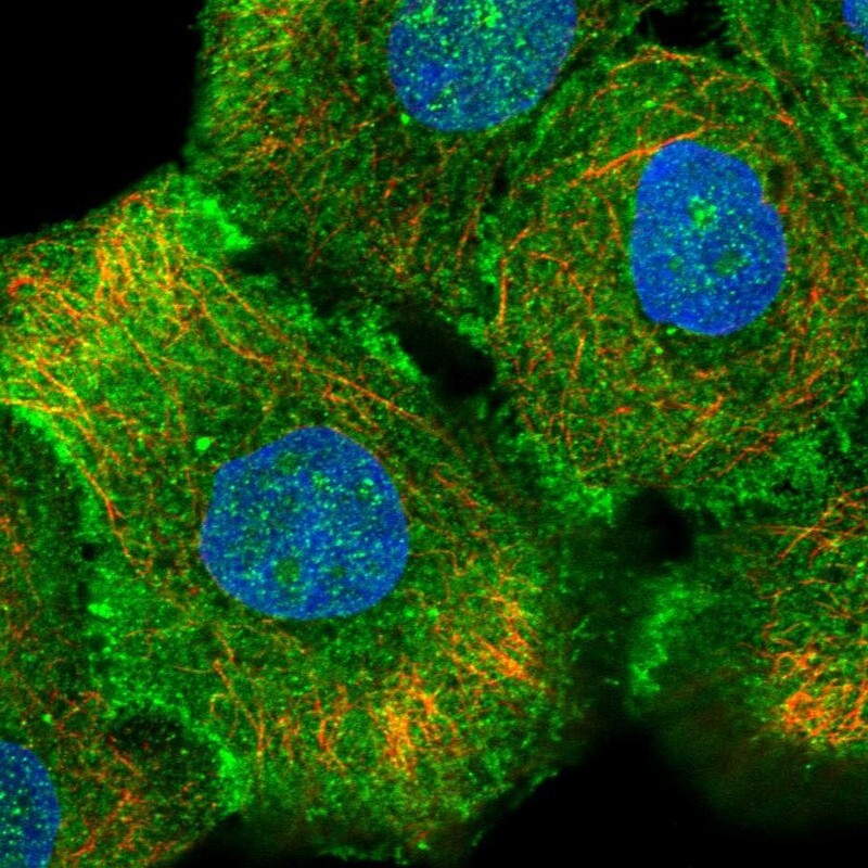
- Experimental details
- Immunofluorescent staining of human cell line HaCaT shows localization to plasma membrane & cell junctions.
- Sample type
- Human