 Explore
 Validate
 Learn
Learn14-1049-37
antibody from Invitrogen Antibodies
Targeting: ITGB4
CD104
 Western blot
 Immunocytochemistry Immunoprecipitation Immunohistochemistry Flow cytometry Other assay
Antibody data
- Antibody Data
- Antigen structure
- References [5]
- Comments [0]
- Validations
- Immunocytochemistry [1]
- Flow cytometry [2]
- Other assay [3]
Submit
Validation data
Reference
Comment
Report error
- Product number
- 14-1049-37 - Provider product page

- Provider
- Invitrogen Antibodies
- Product name
- CD104 (Integrin beta 4) Monoclonal Antibody (439-9B), eBioscience™
- Antibody type
- Monoclonal
- Antigen
- Other
- Description
- Description: The monoclonal antibody 439-9B recognizes human CD104, also known as integrin beta 4. CD104 is a 202 kDa subunit that associates with integrin alpha 6 (CD49f), thereby forming the laminin receptor. Multiple splice variants have been identified. Expression is found on epithelial cells and tumor cells and has been shown to be involved in cell-matrix and cell-cell interactions. The role of CD104 in tumorigenicity is well established. The monoclonal antibody 439-9B has been reported to have no effect on adhesion of human colon adenocarcinoma cells. Moreover, this antibody crossreacts to bovine. Applications Reported: This 439-9B antibody has been reported for use in flow cytometric analysis, immunoprecipitation, immunoblotting under nonreducing conditions only, immunohistology staining of frozen tissue sections, and immunohistology staining of paraffin embedded tissue sections. Applications Tested: This 439-9B antibody has been tested by flow cytometric analysis of A549 cell line. This can be used at less than or equal to 1 µg per test. A test is defined as the amount (µg) of antibody that will stain a cell sample in a final volume of 100 µL. Cell number should be determined empirically but can range from 10^5 to 10^8 cells/test. It is recommended that the antibody be carefully titrated for optimal performance in the assay of interest. Purity: Greater than 90%, as determined by SDS-PAGE. Aggregation: Less than 10%, as determined by HPLC. Filtration: 0.2 µm post-manufacturing filtered.
- Reactivity
- Human
- Host
- Rat
- Isotype
- IgG
- Antibody clone number
- 439-9B
- Vial size
- 2 mg
- Concentration
- 0.5 mg/mL
- Storage
- 4°C
Submitted references Quantitative Proteomics Analysis of Lytic KSHV Infection in Human Endothelial Cells Reveals Targets of Viral Immune Modulation.
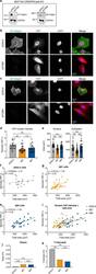
Identification of cell context-dependent YAP-associated proteins reveals β(1) and β(4) integrin mediate YAP translocation independently of cell spreading.
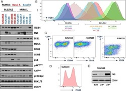
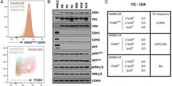
Integrin-β4 identifies cancer stem cell-enriched populations of partially mesenchymal carcinoma cells.
Tumor infiltrating lymphocytes and survival in patients with head and neck squamous cell carcinoma.
Upregulation of integrin β4 promotes epithelial-mesenchymal transition and is a novel prognostic marker in pancreatic ductal adenocarcinoma.
Gabaev I, Williamson JC, Crozier TWM, Schulz TF, Lehner PJ
Cell reports 2020 Oct 13;33(2):108249
Cell reports 2020 Oct 13;33(2):108249
Identification of cell context-dependent YAP-associated proteins reveals β(1) and β(4) integrin mediate YAP translocation independently of cell spreading.
Lee JY, Dominguez AA, Nam S, Stowers RS, Qi LS, Chaudhuri O
Scientific reports 2019 Nov 20;9(1):17188
Scientific reports 2019 Nov 20;9(1):17188
Integrin-β4 identifies cancer stem cell-enriched populations of partially mesenchymal carcinoma cells.
Bierie B, Pierce SE, Kroeger C, Stover DG, Pattabiraman DR, Thiru P, Liu Donaher J, Reinhardt F, Chaffer CL, Keckesova Z, Weinberg RA
Proceedings of the National Academy of Sciences of the United States of America 2017 Mar 21;114(12):E2337-E2346
Proceedings of the National Academy of Sciences of the United States of America 2017 Mar 21;114(12):E2337-E2346
Tumor infiltrating lymphocytes and survival in patients with head and neck squamous cell carcinoma.
Nguyen N, Bellile E, Thomas D, McHugh J, Rozek L, Virani S, Peterson L, Carey TE, Walline H, Moyer J, Spector M, Perim D, Prince M, McLean S, Bradford CR, Taylor JM, Wolf GT, Head and Neck SPORE Program Investigators
Head & neck 2016 Jul;38(7):1074-84
Head & neck 2016 Jul;38(7):1074-84
Upregulation of integrin β4 promotes epithelial-mesenchymal transition and is a novel prognostic marker in pancreatic ductal adenocarcinoma.
Masugi Y, Yamazaki K, Emoto K, Effendi K, Tsujikawa H, Kitago M, Itano O, Kitagawa Y, Sakamoto M
Laboratory investigation; a journal of technical methods and pathology 2015 Mar;95(3):308-19
Laboratory investigation; a journal of technical methods and pathology 2015 Mar;95(3):308-19
No comments: Submit comment
Supportive validation
- Submitted by
- Invitrogen Antibodies (provider)
- Main image
- Experimental details
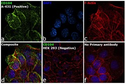
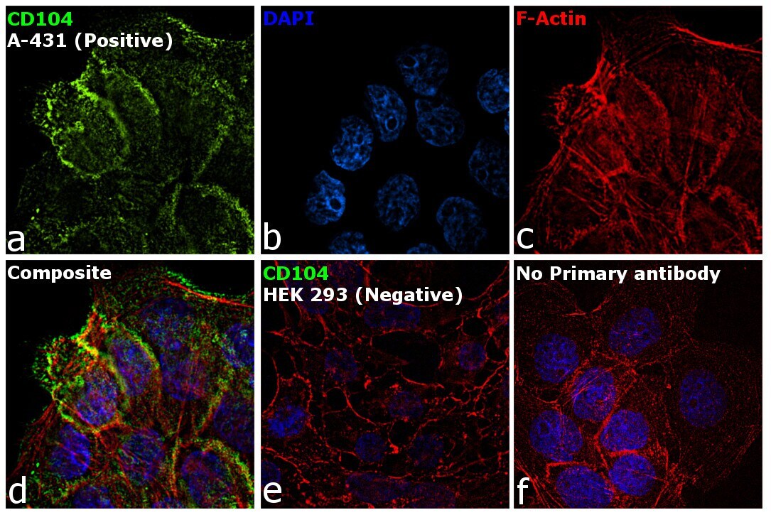
- Immunofluorescence analysis of CD104 (Integrin beta 4) was performed using A-431 and HEK 293 cells. The cells were fixed with 4% paraformaldehyde for 10 minutes, permeabilized with 0.1% Triton™ X-100 for 15 minutes, and blocked with 2% BSA for 1 hour at room temperature. The cells were labeled with P-selectin Mouse Monoclonal Antibody (Product # 14-1049-80) at 5 µg/mL in 0.1% BSA and incubated overnight at 4 degree and then labeled with Goat anti-Rat IgG (H+L) Cross-Adsorbed Secondary Antibody, Alexa Fluor 488 (Product # A-11006) at a dilution of 1:2000 for 45 minutes at room temperature (Panel a: green). Nuclei (Panel b: blue) were stained with ProLong™ Diamond Antifade Mountant with DAPI (Product # P36962). F-actin (Panel c: red) was stained with Rhodamine Phalloidin (Product # R415, 1:300). Panel d represents the composite image showing membrane localization of CD104 in A-431 but not in HEK 293 cells (Panel e). Panel f represents control cells with no primary antibody to assess background. The images were captured at 60X magnification..
Supportive validation
- Submitted by
- Invitrogen Antibodies (provider)
- Main image

- Experimental details
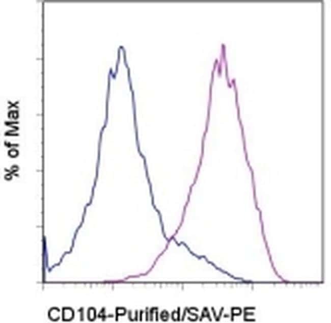
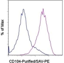
- Staining of the A549 cell line with 0.5 µg of Rat IgG2b K Isotype Control Purified (Product # 14-4031-82) (open histogram) or 0.5 µg of Anti-Human CD104 (Integrin beta 4) Purified (filled histogram) followed by Anti-Rat IgG Biotin (Product # 13-4813-85)and Streptavidin PE (Product # 12-4317-87). Total viable cells were used for analysis.
- Submitted by
- Invitrogen Antibodies (provider)
- Main image
- Experimental details
- Staining of the A549 cell line with 0.5 µg of Rat IgG2b K Isotype Control Purified (Product # 14-4031-82) (open histogram) or 0.5 µg of Anti-Human CD104 (Integrin beta 4) Purified (filled histogram) followed by Anti-Rat IgG Biotin (Product # 13-4813-85)and Streptavidin PE (Product # 12-4317-87). Total viable cells were used for analysis.

Supportive validation
- Submitted by
- Invitrogen Antibodies (provider)
- Main image
- Experimental details
- NULL

- Submitted by
- Invitrogen Antibodies (provider)
- Main image
- Experimental details
- NULL

- Submitted by
- Invitrogen Antibodies (provider)
- Main image
- Experimental details
- Figure 4 beta 1 and beta 4 integrin KO decrease YAP nuclear localization. ( a) Western blot analysis of MCF10A DeltaITGB1 and DeltaITGB4 cells compared to control DeltaGAL4 cells. GAPDH was used as a loading control. Quantification of bands (beta 1 or beta 4 integrin/GAPDH) below each lane. Images cropped from the same blot using different fluorescent intensities. Full-length blot included in Supplementary Fig. 2 . (b) DeltaITGB1 cells stained for beta 1 integrin (green) and YAP (red), DNA stained with DAPI (blue). (c) DeltaITGB4 cells stained for beta 4 integrin (green) and YAP (red), DNA stained with DAPI (blue). (d) YAP nuclear localization in KO cells, **p < 0.01. (e) Nuclear and cytoplasmic areas of KO cells, **p < 0.002. Bars represent mean +- SEM, symbols represent each cell, p-values from one-way ANOVA followed by Tukey post-hoc comparison tests. (f) DeltaGAL4 YAP nuclear localization with area. (g) DeltaITGB1 YAP nuclear localization with area. (h) DeltaITGB4 YAP nuclear localization with area. N = 24-32 cells from 3 independent experiments. (i) Comparison of fits for YAP nuclear localization with area in KO cells. Comparison of (j) Slopes and (k) y-intercepts from best fit lines of YAP nuclear localization with area in KO cells from ( f - h ). **p < 0.01. p-values from linear regression comparing 3 KO best fit lines.
