 Explore
 Validate
 Learn
Learn Western blot
 Immunocytochemistry
Antibody data
- Antibody Data
- Antigen structure
- References [1]
- Comments [0]
- Validations
- Western blot [2]
Submit
Validation data
Reference
Comment
Report error
- Product number
- AF3790 - Provider product page

- Provider
- R&D Systems
- Product name
- Human/Mouse Phospho-SHP-2 (Y542) Antibody
- Antibody type
- Polyclonal
- Description
- Immunogen affinity purified. Detects human and mouse SHP-2 when phosphorylated at Y542.
- Reactivity
- Human, Mouse
- Host
- Rabbit
- Conjugate
- Unconjugated
- Isotype
- IgG
- Vial size
- 100 ug
- Concentration
- LYOPH
- Storage
- Use a manual defrost freezer and avoid repeated freeze-thaw cycles. 12 months from date of receipt, -20 to -70 °C as supplied. 1 month, 2 to 8 °C under sterile conditions after reconstitution. 6 months, -20 to -70 °C under sterile conditions after reconstitution.
Submitted references Helicobacter pylori-related diffuse large B-cell lymphoma of the stomach: a distinct entity with lower aggressiveness and higher chemosensitivity.
Kuo SH, Yeh KH, Chen LT, Lin CW, Hsu PN, Hsu C, Wu MS, Tzeng YS, Tsai HJ, Wang HP, Cheng AL
Blood cancer journal 2014 Jun 20;4(6):e220
Blood cancer journal 2014 Jun 20;4(6):e220
No comments: Submit comment
Supportive validation
- Submitted by
- R&D Systems (provider)
- Main image
- Experimental details
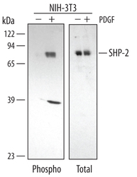
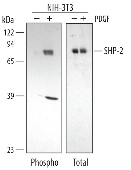
- Detection of Mouse Phospho-SHP-2 (Y542) by Western Blot. Western blot shows lysates of NIH-3T3 mouse embryonic fibroblast cell line untreated (-) or treated (+) with 50 ng/mL Human PDGF-BB (Catalog # 220-BB) for 20 minutes. PVDF membrane was probed with 1 µg/mL of Rabbit Anti-Human/Mouse Phospho-SHP-2 (Y542) Antigen Affinity-purified Polyclonal Antibody (Catalog # AF3790), followed by HRP-conjugated Anti-Rabbit IgG Secondary Antibody (Catalog # HAF008). A specific band was detected for Phospho-SHP-2 (Y542) at approximately 72 kDa (as indicated). The lysates were also probed for total SHP-2 with Human/Mouse/Rat SHP-2 Antigen Affinity-purified Polyclonal Antibody (Catalog # AF1894). This experiment was conducted under reducing conditions and using Immunoblot Buffer Group 1.
- Submitted by
- R&D Systems (provider)
- Main image
- Experimental details
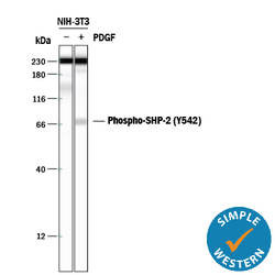
- Detection of Mouse Phospho-SHP-2 (Y542) by Simple WesternTM. Simple Western lane view shows lysates of NIH-3T3 mouse embryonic fibroblast cell line untreated (-) or treated (+) with 50 ng/mL Recombinant Human PDGF-BB (Catalog # 220-BB) for 20 minutes, loaded at 0.2 mg/mL. A specific band was detected for Phospho-SHP-2 (Y542) at approximately 72 kDa (as indicated) using 10 µg/mL of Rabbit Anti-Human/Mouse Phospho-SHP-2 (Y542) Antigen Affinity-purified Polyclonal Antibody (Catalog # AF3790). This experiment was conducted under reducing conditions and using the 12-230 kDa separation system. *Non-specific interaction with the 230 kDa Simple Western standard may be seen with this antibody.
