 Explore
 Validate
 Learn
Learn Western blot
Antibody data
- Antibody Data
- Antigen structure
- References [3]
- Comments [0]
- Validations
- Western blot [2]
- Immunohistochemistry [2]
Submit
Validation data
Reference
Comment
Report error
- Product number
- AF2039 - Provider product page

- Provider
- Novus Biologicals
- Product name
- Goat Polyclonal Smad1 Antibody
- Antibody type
- Polyclonal
- Description
- Antigen Affinity-purified. Detects human Smad1 in direct ELISAs and Western blots. In direct ELISAs, approximately 15% cross-reactivity with recombinant human (rh) Smad5 is observed, and less than 5% cross-reactivity rhSmad4 and rhSmad9 is observed.
- Reactivity
- Human
- Host
- Goat
- Conjugate
- Unconjugated
- Isotype
- IgG
- Vial size
- 100 ug
- Concentration
- LYOPH
- Storage
- Use a manual defrost freezer and avoid repeated freeze-thaw cycles. 12 months from date of receipt, -20 to -70 degreesC as supplied. 1 month, 2 to 8 degreesC under sterile conditions after reconstitution. 6 months, -20 to -70 degreesC under sterile conditions after reconstitution.
Submitted references MicroRNA-155 controls RB phosphorylation in normal and malignant B lymphocytes via the noncanonical TGF-β1/SMAD5 signaling module.
Ca2+/S100 proteins act as upstream regulators of the chaperone-associated ubiquitin ligase CHIP (C terminus of Hsc70-interacting protein).
Fstl1 antagonizes BMP signaling and regulates ureter development.
Jiang D, Aguiar RC
Blood 2014 Jan 2;123(1):86-93
Blood 2014 Jan 2;123(1):86-93
Ca2+/S100 proteins act as upstream regulators of the chaperone-associated ubiquitin ligase CHIP (C terminus of Hsc70-interacting protein).
Shimamoto S, Kubota Y, Yamaguchi F, Tokumitsu H, Kobayashi R
The Journal of biological chemistry 2013 Mar 8;288(10):7158-68
The Journal of biological chemistry 2013 Mar 8;288(10):7158-68
Fstl1 antagonizes BMP signaling and regulates ureter development.
Xu J, Qi X, Gong J, Yu M, Zhang F, Sha H, Gao X
PloS one 2012;7(4):e32554
PloS one 2012;7(4):e32554
No comments: Submit comment
Supportive validation
- Submitted by
- Novus Biologicals (provider)
- Main image
- Experimental details
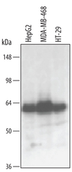
- Detection of Human Smad1 by Western Blot. Western blot shows lysates of HepG2 human hepatocellular carcinoma cell line, MBA-MB-468 human breast cancer cell line, and HT-29 human colon adenocarcinoma cell line. PVDF membrane was probed with 0.5 µg/mL of Goat Anti-Human Smad1 Antigen Affinity-purified Polyclonal Antibody (Catalog # AF2039) followed by HRP-conjugated Anti-Goat IgG Secondary Antibody (Catalog # HAF109). A specific band was detected for Smad1 at approximately 63 kDa (as indicated). This experiment was conducted under reducing conditions and using Immunoblot Buffer Group 1.

- Submitted by
- Novus Biologicals (provider)
- Main image
- Experimental details
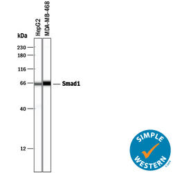
- Detection of Human Smad1 by Simple WesternTM. Simple Western lane view shows lysates of HepG2 human hepatocellular carcinoma cell line and MDA-MB-468 human breast cancer cell line, loaded at 0.2 mg/mL. A specific band was detected for Smad1 at approximately 66 kDa (as indicated) using 5 µg/mL of Goat Anti-Human Smad1 Antigen Affinity-purified Polyclonal Antibody (Catalog # AF2039) followed by 1:50 dilution of HRP-conjugated Anti-Goat IgG Secondary Antibody (Catalog # HAF109). This experiment was conducted under reducing conditions and using the 12-230 kDa separation system.

Supportive validation
- Submitted by
- Novus Biologicals (provider)
- Main image

- Experimental details
- Smad1 in Human Breast. Smad1 was detected in immersion fixed paraffin-embedded sections of human breast array using Goat Anti-Human Smad1 Antigen Affinity-purified Polyclonal Antibody (Catalog # AF2039) at 15 µg/mL overnight at 4 °C. Tissue was stained using the Anti-Goat HRP-DAB Cell & Tissue Staining Kit (brown; Catalog # CTS008) and counterstained with hematoxylin (blue). Lower panel shows a lack of labeling if primary antibodies are omitted and tissue is stained only with secondary antibody followed by incubation with detection reagents. View our protocol for Chromogenic IHC Staining of Paraffin-embedded Tissue Sections.

- Submitted by
- Novus Biologicals (provider)
- Main image

- Experimental details
- Smad1 in Human Breast Cancer Tissue. Smad1 was detected in immersion fixed paraffin-embedded sections of human breast cancer tissue using Goat Anti-Human Smad1 Antigen Affinity-purified Polyclonal Antibody (Catalog # AF2039) at 15 µg/mL overnight at 4 °C. Tissue was stained using the Anti-Goat HRP-DAB Cell & Tissue Staining Kit (brown; Catalog # CTS008) and counterstained with hematoxylin (blue). Specific labeling was localized to the nuclei of glandular epithelial cells. View our protocol for Chromogenic IHC Staining of Paraffin-embedded Tissue Sections.
