Explore
 Validate
 Learn
Learn Western blot
 Immunoprecipitation
Antibody data
- Antibody Data
- Antigen structure
- References [1]
- Comments [0]
- Validations
- Western blot [3]
Submit
Validation data
Reference
Comment
Report error
- Product number
- PA5-17122 - Provider product page

- Provider
- Invitrogen Antibodies
- Product name
- SMAD1 Polyclonal Antibody
- Antibody type
- Polyclonal
- Antigen
- Synthetic peptide
- Description
- It is not recommended to aliquot this antibody.
- Reactivity
- Human, Mouse
- Host
- Rabbit
- Isotype
- IgG
- Vial size
- 100 µL
- Concentration
- 83 µg/mL
- Storage
- -20°C
Submitted references Inhibition of osteoblastic Smurf1 promotes bone formation in mouse models of distinctive age-related osteoporosis.
Liang C, Peng S, Li J, Lu J, Guan D, Jiang F, Lu C, Li F, He X, Zhu H, Au DWT, Yang D, Zhang BT, Lu A, Zhang G
Nature communications 2018 Aug 24;9(1):3428
Nature communications 2018 Aug 24;9(1):3428
No comments: Submit comment
Supportive validation
- Submitted by
- Invitrogen Antibodies (provider)
- Main image
- Experimental details
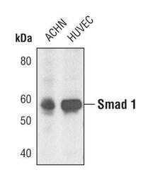
- Western blot analysis of Smad1 in extracts from ACHN and HUVEC cell lines using Smad1 polyclonal antibody (Product # PA5-17122).
- Submitted by
- Invitrogen Antibodies (provider)
- Main image

- Experimental details
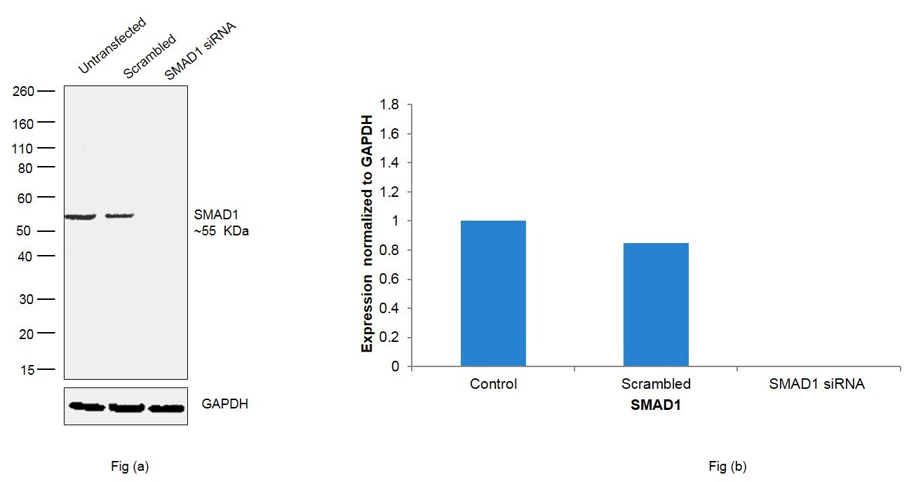
- Knockdown of SMAD1 was achieved by transfecting T-47D cells with SMAD1 specific siRNAs (Silencer® select Product # s8395, s8394). Western blot analysis (Fig. a) was performed using whole cell extracts from the SMAD1 knockdown cells (lane 3), non-specific scrambled siRNA transfected cells (lane 2) and untransfected cells (lane 1). The blot was probed with SMAD1 Polyclonal Antibody (Product # PA5-17122, 1:1000 dilution) and Goat anti-Rabbit IgG (H+L), Superclonal™ Recombinant Secondary Antibody, HRP (Product # A27036, 1:4000 dilution) . Densitometric analysis of this western blot is shown in histogram (Fig. b). Decrease in signal upon siRNA mediated knock down confirms that antibody is specific to SMAD1.
- Submitted by
- Invitrogen Antibodies (provider)
- Main image
- Experimental details
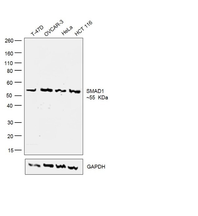
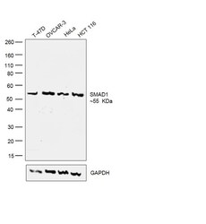
- Western blot was performed using anti-SMAD1 Polyclonal Antibody (Product # PA5-17122) and a 55 kDa band corresponding to SMAD1 was observed across the cell lines tested. Modified Whole cell extracts (30 µg lysate) (1% SDS) of T-47D (Lane 1), OVCAR-3 (Lane 2), HeLa (Lane 3) and HCT 116 (Lane 4) were electrophoresed using Novex® NuPAGE® 4-12 % Bis-Tris gel (Product # NP0322BOX). Resolved proteins were then transferred onto a nitrocellulose membrane (Product # IB23001) by iBlot® 2 Dry Blotting System (Product # IB21001). The blot was probed with the primary antibody (1:1000 dilution) and detected by chemiluminescence with Goat anti-Rabbit IgG (H+L) Superclonal™ Secondary Antibody, HRP (Product # A27036, 1:4000 dilution) using the iBright FL 1000 (Product # A32752). Chemiluminescent detection was performed using Novex® ECL Chemiluminescent Substrate Reagent Kit (Product # WP20005).