 Explore
 Validate
 Learn
Learn Western blot
 Immunocytochemistry
Antibody data
- Antibody Data
- Antigen structure
- References [2]
- Comments [0]
- Validations
- Western blot [2]
- Flow cytometry [4]
Submit
Validation data
Reference
Comment
Report error
- Product number
- NBP2-27369 - Provider product page

- Provider
- Novus Biologicals
- Product name
- Mouse Monoclonal MyD88 Antibody
- Antibody type
- Monoclonal
- Description
- Protein G purified. The immunogen is 100% homologous to isoform 3: 264 aa, isoform 4: 204 aa
- Reactivity
- Human, Mouse, Simian
- Host
- Mouse
- Isotype
- IgG
- Vial size
- 0.1 mg
- Concentration
- 1.0 mg/ml
- Storage
- Store at 4C short term. Aliquot and store at -20C long term. Avoid freeze-thaw cycles.
Submitted references Early Differentiation of Human CD11c+NK Cells with γδ T Cell Activation Properties Is Promoted by Dialyzable Leukocyte Extracts.
Microglia Induce Neurotoxic IL-17+ γδ T Cells Dependent on TLR2, TLR4, and TLR9 Activation.
Ramírez-Ramírez D, Vadillo E, Arriaga-Pizano LA, Mayani H, Estrada-Parra S, Velasco-Velázquez MA, Pérez-Tapia SM, Pelayo R
Journal of immunology research 2016;2016:4097642
Journal of immunology research 2016;2016:4097642
Microglia Induce Neurotoxic IL-17+ γδ T Cells Dependent on TLR2, TLR4, and TLR9 Activation.
Derkow K, Krüger C, Dembny P, Lehnardt S
PloS one 2015;10(8):e0135898
PloS one 2015;10(8):e0135898
No comments: Submit comment
Supportive validation
- Submitted by
- Novus Biologicals (provider)
- Main image

- Experimental details
- Western Blot: MyD88 Antibody (4D6) [NBP2-27369] - Analysis of MyD88 in A) human ovary, B) human prostate and Jurkat cell lysate in the C) absence and D) presence of immunizing peptide using this antibody. Goat anti-mouse Ig HRP secondary antibody and PicoTect ECL substrate solution were used for this test.

- Submitted by
- Novus Biologicals (provider)
- Main image

- Experimental details
- Western Blot: MyD88 Antibody (4D6) [NBP2-27369] - Normal human bronchial epithelial total cell lysate. Image from verified customer review.

Supportive validation
- Submitted by
- Novus Biologicals (provider)
- Main image

- Experimental details
- Flow Cytometry: MyD88 Antibody (4D6) [NBP2-27369] - Intracellular analysis of MyD88 antibody in Jurkat cells using 0.5 ug. Shaded histogram represents cells without antibody; green represents isotype control; red represents MyD88 antibody. This antibody was used for this test, and an anti-mouse IgG PE conjugated secondary antibody.

- Submitted by
- Novus Biologicals (provider)
- Main image

- Experimental details
- Flow (Intracellular): MyD88 Antibody (4D6) [NBP2-27369] - Analysis using the Alexa Fluor (R) 488 conjugate of NBP2-27369. Staining of MyD88 antibody in Jurkat cells using 10 ul (0.5 ug) of MyD88 antibody. Green represents isotype control (20108); red represents anti-MyD88 antibody.

- Submitted by
- Novus Biologicals (provider)
- Main image

- Experimental details
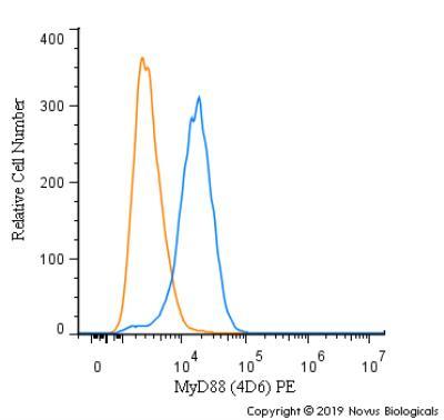
- Flow Cytometry: MyD88 Antibody (4D6) [NBP2-27369] - An intracellular stain was performed on MCF7 cells with MyD88 (4D6) antibody NBP2-27369PE (blue) and a matched isotype control (orange). Cells were fixed with 4% PFA and then permeablized with 0.1% saponin. Cells were incubated in an antibody dilution of 2.5 ug/mL for 30 minutes at room temperature. Both antibodies were conjugated to Phycoerythrin.
- Submitted by
- Novus Biologicals (provider)
- Main image

- Experimental details
- Flow Cytometry: MyD88 Antibody (4D6) [NBP2-27369] - An intracellular stain was performed on MCF7 cells with MyD88 [4D6] Antibody NBP2-27369AF488 (blue) and a matched isotype control (orange). Cells were fixed with 4% PFA and then permeabilized with 0.1% saponin. Cells were incubated in an antibody dilution of 10 ug/mL for 30 minutes at room temperature. Both antibodies were conjugated to Alexa Fluor 488.
