Explore
 Validate
 Learn
Learn Western blot
Antibody data
- Antibody Data
- Antigen structure
- References [5]
- Comments [0]
- Validations
- Western blot [3]
- Immunocytochemistry [1]
- Flow cytometry [1]
Submit
Validation data
Reference
Comment
Report error
- Product number
- AF3109 - Provider product page

- Provider
- R&D Systems
- Product name
- Mouse/Rat MyD88 Antibody
- Antibody type
- Polyclonal
- Description
- Antigen Affinity-purified. Detects mouse and rat MyD88 in Western blots.
- Reactivity
- Mouse, Rat
- Host
- Goat
- Conjugate
- Unconjugated
- Antigen sequence
Q3U7M4- Isotype
- IgG
- Vial size
- 100 ug
- Concentration
- LYOPH
- Storage
- Use a manual defrost freezer and avoid repeated freeze-thaw cycles. 12 months from date of receipt, -20 to -70 °C as supplied. 1 month, 2 to 8 °C under sterile conditions after reconstitution. 6 months, -20 to -70 °C under sterile conditions after reconstitution.
Submitted references The Single Nucleotide Polymorphism Mal-D96N Mice Provide New Insights into Functionality of Mal in TLR Immune Responses.
Interleukin-1 receptor-associated kinase 4 (IRAK4) plays a dual role in myddosome formation and Toll-like receptor signaling.
Blocking TIR Domain Interactions in TLR9 Signaling.
Signaling through the adaptor molecule MyD88 in CD4+ T cells is required to overcome suppression by regulatory T cells.
Toll-like receptor 2-mediated signaling requirements for Francisella tularensis live vaccine strain infection of murine macrophages.
Dowling JK, Tate MD, Rosli S, Bourke NM, Bitto N, Lauterbach MA, Cheung S, Ve T, Kobe B, Golenbock D, Mansell A
Journal of immunology (Baltimore, Md. : 1950) 2019 Apr 15;202(8):2384-2396
Journal of immunology (Baltimore, Md. : 1950) 2019 Apr 15;202(8):2384-2396
Interleukin-1 receptor-associated kinase 4 (IRAK4) plays a dual role in myddosome formation and Toll-like receptor signaling.
De Nardo D, Balka KR, Cardona Gloria Y, Rao VR, Latz E, Masters SL
The Journal of biological chemistry 2018 Sep 28;293(39):15195-15207
The Journal of biological chemistry 2018 Sep 28;293(39):15195-15207
Blocking TIR Domain Interactions in TLR9 Signaling.
Javmen A, Szmacinski H, Lakowicz JR, Toshchakov VY
Journal of immunology (Baltimore, Md. : 1950) 2018 Aug 1;201(3):995-1006
Journal of immunology (Baltimore, Md. : 1950) 2018 Aug 1;201(3):995-1006
Signaling through the adaptor molecule MyD88 in CD4+ T cells is required to overcome suppression by regulatory T cells.
Schenten D, Nish SA, Yu S, Yan X, Lee HK, Brodsky I, Pasman L, Yordy B, Wunderlich FT, Brüning JC, Zhao H, Medzhitov R
Immunity 2014 Jan 16;40(1):78-90
Immunity 2014 Jan 16;40(1):78-90
Toll-like receptor 2-mediated signaling requirements for Francisella tularensis live vaccine strain infection of murine macrophages.
Cole LE, Shirey KA, Barry E, Santiago A, Rallabhandi P, Elkins KL, Puche AC, Michalek SM, Vogel SN
Infection and immunity 2007 Aug;75(8):4127-37
Infection and immunity 2007 Aug;75(8):4127-37
No comments: Submit comment
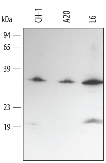
Supportive validation
- Submitted by
- R&D Systems (provider)
- Main image
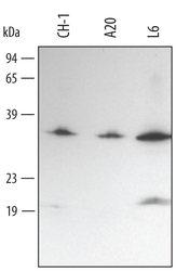
- Experimental details
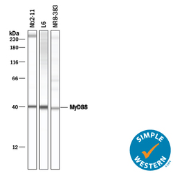
- Detection of Mouse/Rat MyD88 by Western Blot. Western blot shows lysates of L6 rat myoblast cell line, A20 mouse B cell lymphoma cell line, and CH-1 mouse B cell lymphoma cell line. PVDF membrane was probed with 1 µg/mL of Goat Anti-Mouse/Rat MyD88 Antigen Affinity-purified Polyclonal Antibody (Catalog # AF3109) followed by HRP-conjugated Anti-Goat IgG Secondary Antibody (Catalog # HAF017). A specific band was detected for MyD88 at approximately 34 kDa (as indicated). This experiment was conducted under reducing conditions and using Immunoblot Buffer Group 2.
- Submitted by
- R&D Systems (provider)
- Main image
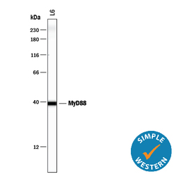
- Experimental details
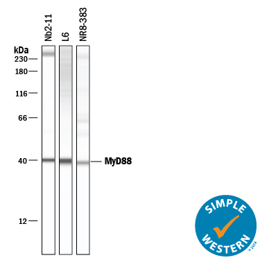
- Detection of Rat MyD88 by Simple WesternTM. Simple Western lane view shows lysates of L6 rat myoblast cell line, loaded at 0.2 mg/mL. A specific band was detected for MyD88 at approximately 39 kDa (as indicated) using 10 µg/mL of Goat Anti-Mouse/Rat MyD88 Antigen Affinity-purified Polyclonal Antibody (Catalog # AF3109) followed by 1:50 dilution of HRP-conjugated Anti-Goat IgG Secondary Antibody (Catalog # HAF109). This experiment was conducted under reducing conditions and using the 12-230 kDa separation system.

- Submitted by
- R&D Systems (provider)
- Main image
- Experimental details
- Detection of Rat MyD88 by Simple WesternTM. Simple Western lane view shows lysates of L6 rat myoblast cell line, loaded at 0.2 mg/mL. A specific band was detected for MyD88 at approximately 39 kDa (as indicated) using 10 µg/mL of Goat Anti-Mouse/Rat MyD88 Antigen Affinity-purified Polyclonal Antibody (Catalog # AF3109) followed by 1:50 dilution of HRP-conjugated Anti-Goat IgG Secondary Antibody (Catalog # HAF109). This experiment was conducted under reducing conditions and using the 12-230 kDa separation system.
Supportive validation
- Submitted by
- R&D Systems (provider)
- Main image

- Experimental details
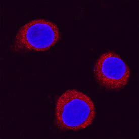
- MyD88 in RAW 264.7 Mouse Cell Line. MyD88 was detected in immersion fixed RAW 264.7 mouse monocyte/macrophage cell line using Goat Anti-Mouse/Rat MyD88 Antigen Affinity-purified Polyclonal Antibody (Catalog # AF3109) at 1.7 µg/mL for 3 hours at room temperature. Cells were stained using the NorthernLights™ 557-conjugated Anti-Goat IgG Secondary Antibody (red; Catalog # NL001) and counterstained with DAPI(blue). Specific staining was localized to cytoplasmic. View our protocol for Fluorescent ICC Staining of Cells on Coverslips.
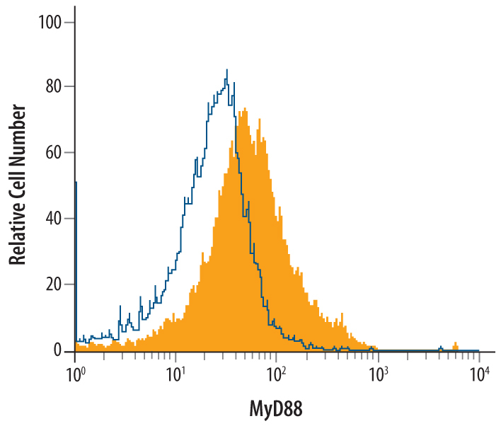
Supportive validation
- Submitted by
- R&D Systems (provider)
- Main image
- Experimental details
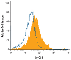
- Detection of MyD88 in Mouse Splenocytes by Flow Cytometry. Mouse splenocytes were stained with Goat Anti-Mouse/Rat MyD88 Antigen Affinity-purified Polyclonal Antibody (Catalog # AF3109, filled histogram) or control antibody (Catalog # AB-108-C, open histogram), followed by Allophycocyanin-conjugated Anti-Goat IgG Secondary Antibody (Catalog # F0108). To facilitate intracellular staining, cells were fixed with paraformaldehyde and permeabilized with saponin. This application has not been tested in rat samples.