 Explore
 Validate
 Learn
Learn Immunocytochemistry
Antibody data
- Antibody Data
- Antigen structure
- References [2]
- Comments [0]
- Validations
- Immunocytochemistry [1]
Submit
Validation data
Reference
Comment
Report error
- Product number
- MAB3109 - Provider product page

- Provider
- R&D Systems
- Product name
- Mouse MyD88 Antibody
- Antibody type
- Monoclonal
- Description
- Protein A or G purified from hybridoma culture supernatant. Detects mouse MyD88 in direct ELISAs.
- Reactivity
- Mouse
- Host
- Rat
- Conjugate
- Unconjugated
- Antigen sequence
Q3U7M4- Isotype
- IgG
- Antibody clone number
- 316902
- Vial size
- 100 ug
- Concentration
- LYOPH
- Storage
- Use a manual defrost freezer and avoid repeated freeze-thaw cycles. 12 months from date of receipt, -20 to -70 °C as supplied. 1 month, 2 to 8 °C under sterile conditions after reconstitution. 6 months, -20 to -70 °C under sterile conditions after reconstitution.
Submitted references HMGB1 is an early and critical mediator in an animal model of uveitis induced by IRBP-specific T cells.
Bacterial recognition by TLR7 in the lysosomes of conventional dendritic cells.
Jiang G, Sun D, Yang H, Lu Q, Kaplan HJ, Shao H
Journal of leukocyte biology 2014 Apr;95(4):599-607
Journal of leukocyte biology 2014 Apr;95(4):599-607
Bacterial recognition by TLR7 in the lysosomes of conventional dendritic cells.
Mancuso G, Gambuzza M, Midiri A, Biondo C, Papasergi S, Akira S, Teti G, Beninati C
Nature immunology 2009 Jun;10(6):587-94
Nature immunology 2009 Jun;10(6):587-94
No comments: Submit comment
Supportive validation
- Submitted by
- R&D Systems (provider)
- Main image
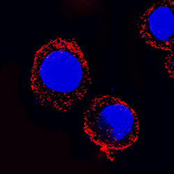
- Experimental details
- MyD88 in RAW 264.7 Mouse Cell Line. MyD88 was detected in immersion fixed RAW 264.7 mouse monocyte/macrophage cell line using Rat Anti-Mouse MyD88 Monoclonal Antibody (Catalog # MAB3109) at 25 µg/mL for 3 hours at room temperature. Cells were stained using the NorthernLights™ 557-conjugated Anti-Rat IgG Secondary Antibody (red; Catalog # NL014) and counterstained with DAPI (blue). Specific staining was localized to cytoplasm. View our protocol for Fluorescent ICC Staining of Non-adherent Cells.
