Explore
 Validate
 Learn
Learn Western blot
Antibody data
- Antibody Data
- Antigen structure
- References [3]
- Comments [0]
- Validations
- Western blot [2]
- Immunocytochemistry [1]
- Flow cytometry [1]
Submit
Validation data
Reference
Comment
Report error
- Product number
- AF2928 - Provider product page

- Provider
- R&D Systems
- Product name
- Human MyD88 Antibody
- Antibody type
- Polyclonal
- Description
- Antigen Affinity-purified. Detects human MyD88 in direct ELISAs and Western blots. In direct ELISAs, approximately 20% cross-reactivity with recombinant mouse MyD88 is observed.
- Reactivity
- Human
- Host
- Goat
- Conjugate
- Unconjugated
- Antigen sequence
Q99836- Isotype
- IgG
- Vial size
- 100 ug
- Concentration
- LYOPH
- Storage
- Use a manual defrost freezer and avoid repeated freeze-thaw cycles. 12 months from date of receipt, -20 to -70 °C as supplied. 1 month, 2 to 8 °C under sterile conditions after reconstitution. 6 months, -20 to -70 °C under sterile conditions after reconstitution.
Submitted references Cell-Based Screen Identifies Human Interferon-Stimulated Regulators of Listeria monocytogenes Infection.
Enterovirus 68 3C protease cleaves TRIF to attenuate antiviral responses mediated by Toll-like receptor 3.
Potentiation of flagellin responses in gut epithelial cells by interferon-gamma is associated with STAT-independent regulation of MyD88 expression.
Perelman SS, Abrams ME, Eitson JL, Chen D, Jimenez A, Mettlen M, Schoggins JW, Alto NM
PLoS pathogens 2016 Dec;12(12):e1006102
PLoS pathogens 2016 Dec;12(12):e1006102
Enterovirus 68 3C protease cleaves TRIF to attenuate antiviral responses mediated by Toll-like receptor 3.
Xiang Z, Li L, Lei X, Zhou H, Zhou Z, He B, Wang J
Journal of virology 2014 Jun;88(12):6650-9
Journal of virology 2014 Jun;88(12):6650-9
Potentiation of flagellin responses in gut epithelial cells by interferon-gamma is associated with STAT-independent regulation of MyD88 expression.
Bannon C, Davies PJ, Collett A, Warhurst G
The Biochemical journal 2009 Sep 14;423(1):119-28
The Biochemical journal 2009 Sep 14;423(1):119-28
No comments: Submit comment
Supportive validation
- Submitted by
- R&D Systems (provider)
- Main image
- Experimental details
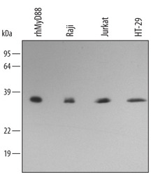
- Detection of Human MyD88 by Western Blot. Western blot shows lysates of Raji human Burkitt's lymphoma cell line, Jurkat human acute T cell leukemia cell line, and HT-29 human colon adenocarcinoma cell line. PVDF membrane was probed with 0.5 µg/mL Goat Anti-Human MyD88 Antigen Affinity-purified Polyclonal Antibody (Catalog # AF2928) followed by HRP-conjugated Anti-Goat IgG Secondary Antibody (Catalog # HAF017). For additional reference, recombinant human MyD88 (1 ng) was included. A specific band for MyD88 was detected at approximately 35 kDa (as indicated). This experiment was conducted under reducing conditions and using Immunoblot Buffer Group 3.
- Submitted by
- R&D Systems (provider)
- Main image

- Experimental details
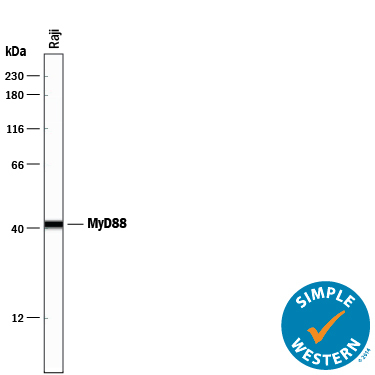
- Detection of Human MyD88 by Simple WesternTM. Simple Western lane view shows lysates of Raji human Burkitt's lymphoma cell line, loaded at 0.2 mg/mL. A specific band was detected for MyD88 at approximately 41 kDa (as indicated) using 5 µg/mL of Goat Anti-Human MyD88 Antigen Affinity-purified Polyclonal Antibody (Catalog # AF2928) followed by 1:50 dilution of HRP-conjugated Anti-Goat IgG Secondary Antibody (Catalog # HAF109). This experiment was conducted under reducing conditions and using the 12-230 kDa separation system.
Supportive validation
- Submitted by
- R&D Systems (provider)
- Main image

- Experimental details
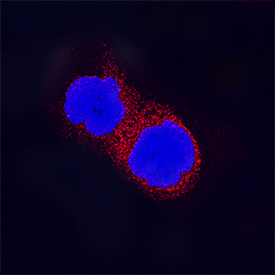
- MyD88 in Raji Human Cell Line. MyD88 was detected in immersion fixed Raji human Burkitt's lymphoma cell line using Goat Anti-Human MyD88 Antigen Affinity-purified Polyclonal Antibody (Catalog # AF2928) at 15 µg/mL for 3 hours at room temperature. Cells were stained using the NorthernLights™ 557-conjugated Anti-Goat IgG Secondary Antibody (red; Catalog # NL001) and counterstained with DAPI (blue). Specific staining was localized to cytoplasm. View our protocol for Fluorescent ICC Staining of Non-adherent Cells.
Supportive validation
- Submitted by
- R&D Systems (provider)
- Main image

- Experimental details
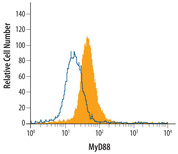
- Detection of MyD88 in Raji Human Cell Line by Flow Cytometry. Raji human Burkitt's lymphoma cell line was stained with Goat Anti-Human MyD88 Antigen Affinity-purified Polyclonal Antibody (Catalog # AF2928, filled histogram) or isotype control antibody (Catalog # AB-108-C, open histogram), followed by Allophycocyanin-conjugated Anti-Goat IgG Secondary Antibody (Catalog # F0108). To facilitate intracellular staining, cells were fixed with paraformaldehyde and permeabilized with saponin.