Explore
 Validate
 Learn
Learn Western blot
Antibody data
- Antibody Data
- Antigen structure
- References [5]
- Comments [0]
- Validations
- Western blot [3]
- Immunocytochemistry [3]
- Immunohistochemistry [1]
- Other assay [4]
Submit
Validation data
Reference
Comment
Report error
- Product number
- PA5-29253 - Provider product page

- Provider
- Invitrogen Antibodies
- Product name
- XIAP Polyclonal Antibody
- Antibody type
- Polyclonal
- Antigen
- Recombinant full-length protein
- Description
- Recommended positive controls: HeLa, Mouse brain. Predicted reactivity: Mouse (90%), Rat (88%), Dog (85%), Pig (89%), Rabbit (89%), Bovine (87%). Store product as a concentrated solution. Centrifuge briefly prior to opening the vial.
- Reactivity
- Human, Mouse
- Host
- Rabbit
- Isotype
- IgG
- Vial size
- 100 μL
- Concentration
- 1 mg/mL
- Storage
- Store at 4°C short term. For long term storage, store at -20°C, avoiding freeze/thaw cycles.
Submitted references Suppression of EGFR/PKC-δ/NF-κB Signaling Associated With Imipramine-Inhibited Progression of Non-Small Cell Lung Cancer.
HtrA4 Protease Promotes Chemotherapeutic-Dependent Cancer Cell Death.
Protein Kinase B and Extracellular Signal-Regulated Kinase Inactivation is Associated with Regorafenib-Induced Inhibition of Osteosarcoma Progression In Vitro and In Vivo.
Fluoxetine Induces Apoptosis through Extrinsic/Intrinsic Pathways and Inhibits ERK/NF-κB-Modulated Anti-Apoptotic and Invasive Potential in Hepatocellular Carcinoma Cells In Vitro.
Magnolol Induces Apoptosis and Inhibits ERK-modulated Metastatic Potential in Hepatocellular Carcinoma Cells.
Yueh PF, Lee YH, Chiang IT, Chen WT, Lan KL, Chen CH, Hsu FT
Frontiers in oncology 2021;11:735183
Frontiers in oncology 2021;11:735183
HtrA4 Protease Promotes Chemotherapeutic-Dependent Cancer Cell Death.
Wenta T, Rychlowski M, Jarzab M, Lipinska B
Cells 2019 Sep 20;8(10)
Cells 2019 Sep 20;8(10)
Protein Kinase B and Extracellular Signal-Regulated Kinase Inactivation is Associated with Regorafenib-Induced Inhibition of Osteosarcoma Progression In Vitro and In Vivo.
Pan PJ, Liu YC, Hsu FT
Journal of clinical medicine 2019 Jun 24;8(6)
Journal of clinical medicine 2019 Jun 24;8(6)
Fluoxetine Induces Apoptosis through Extrinsic/Intrinsic Pathways and Inhibits ERK/NF-κB-Modulated Anti-Apoptotic and Invasive Potential in Hepatocellular Carcinoma Cells In Vitro.
Chen WT, Hsu FT, Liu YC, Chen CH, Hsu LC, Lin SS
International journal of molecular sciences 2019 Feb 11;20(3)
International journal of molecular sciences 2019 Feb 11;20(3)
Magnolol Induces Apoptosis and Inhibits ERK-modulated Metastatic Potential in Hepatocellular Carcinoma Cells.
Kuan LY, Chen WL, Chen JH, Hsu FT, Liu TT, Chen WT, Wang KL, Chen WC, Liu YC, Wang WS
In vivo (Athens, Greece) 2018 Nov-Dec;32(6):1361-1368
In vivo (Athens, Greece) 2018 Nov-Dec;32(6):1361-1368
No comments: Submit comment
Supportive validation
- Submitted by
- Invitrogen Antibodies (provider)
- Main image

- Experimental details
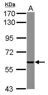
- Western Blot using XIAP Polyclonal Antibody (Product # PA5-29253). Sample (50 µg of whole cell lysate). Lane A: Mouse brain . 7.5% SDS PAGE. XIAP Polyclonal Antibody (Product # PA5-29253) diluted at 1:1,000.
- Submitted by
- Invitrogen Antibodies (provider)
- Main image
- Experimental details
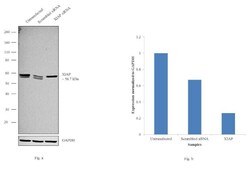
- Knockdown of XIAP was achieved by transfecting HeLa cells with XIAP specific siRNAs (Silencer® select Product # s1455). Western blot analysis (Fig. a) was performed using whole cell extracts from the XIAP knockdown cells (lane 3), non-specific scrambled siRNA transfected cells (lane 2) and untransfected cells (lane 1). The blots were probed with Anti-XIAP Polyclonal Antibody (Product # PA5-29253, 1:2000 dilution) and Goat anti-Rabbit IgG (Heavy Chain) Superclonal™ Secondary Antibody, HRP conjugate (Product # A27036, 0.25 µg/mL, 1:4000 dilution). Knock down was observed for the 56.7 kDa band corresponding to the XIAP canonical isoform as shown in the histogram post densitometric analysis (Fig. b). The ~60 kDa band corresponding to XIAP isoform X1 remained unaffected as the siRNA used specifically targets the XIAP canonical form. Decrease in signal upon siRNA mediated knock down confirms that antibody is specific to XIAP

- Submitted by
- Invitrogen Antibodies (provider)
- Main image
- Experimental details
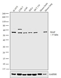
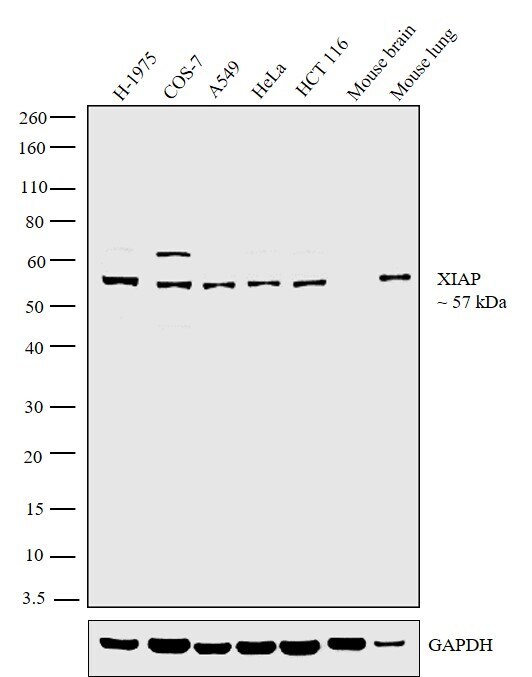
- Western blot analysis was performed on whole cell extracts (30 µg lysate) of H-1975 (Lane 1), COS-7 (Lane 2), A549 (Lane 3), HeLa (Lane 4), HCT 116 (Lane 5), Mouse brain (Lane 6) and Mouse lung (Lane 7). The blot was probed with Anti-XIAP Polyclonal Antibody (Product # PA5-29253, 1:1000 dilution) and detected by chemiluminescence using Goat anti-Rabbit IgG (Heavy Chain) Superclonal™ Secondary Antibody, HRP conjugate (Product # A27036, 0.25 µg/mL, 1:4000 dilution). A 57 kDa band corresponding to XIAP was observed across the cell lines tested (Lanes 1-5). However, the band was absent in Mouse brain which is reported negative for XIAP expression (Lane 6), while it was present in Mouse lung (Lane 7).
Supportive validation
- Submitted by
- Invitrogen Antibodies (provider)
- Main image

- Experimental details
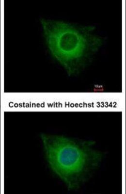
- Immunofluorescent analysis of XIAP in methanol-fixed HeLa cells using a XIAP polyclonal antibody (Product # PA5-29253) at a 1:500 dilution.
- Submitted by
- Invitrogen Antibodies (provider)
- Main image

- Experimental details
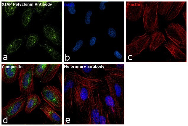
- Immunofluorescence analysis of XIAP was performed using 70% confluent log phase HeLa cells. The cells were fixed with 4% paraformaldehyde for 10 minutes, permeabilized with 0.1% Triton™ X-100 for 15 minutes, and blocked with 1% BSA for 1 hour at room temperature. The cells were labeled with anti-XIAP Rabbit Polyclonal Antibody (Product # PA5-29253) at 1:200 dilution in 0.1% BSA, incubated at 4 degree Celsius overnight and then labeled with Goat anti-Rabbit IgG (H+L) Superclonal™ Secondary Antibody, Alexa Fluor® 488 conjugate (Product # A27034) at a dilution of 1:2000 for 45 minutes at room temperature (Panel a: green). Nuclei (Panel b: blue) were stained with ProLong™ Diamond Antifade Mountant with DAPI (Product # P36962). F-actin (Panel c: red) was stained with Rhodamine Phalloidin (Product # R415, 1:300). Panel d represents the merged image showing nuclear and cytoplasmic localization. Panel e represents control cells with no primary antibody to assess background. The images were captured at 60X magnification.
- Submitted by
- Invitrogen Antibodies (provider)
- Main image
- Experimental details
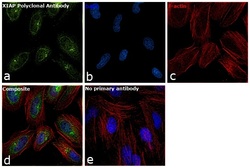
- Immunofluorescence analysis of XIAP was performed using 70% confluent log phase HeLa cells. The cells were fixed with 4% paraformaldehyde for 10 minutes, permeabilized with 0.1% Triton™ X-100 for 15 minutes, and blocked with 1% BSA for 1 hour at room temperature. The cells were labeled with anti-XIAP Rabbit Polyclonal Antibody (Product # PA5-29253) at 1:200 dilution in 0.1% BSA, incubated at 4 degree Celsius overnight and then labeled with Goat anti-Rabbit IgG (Heavy Chain) Superclonal™ Secondary Antibody, Alexa Fluor® 488 conjugate (Product # A27034) at a dilution of 1:2000 for 45 minutes at room temperature (Panel a: green). Nuclei (Panel b: blue) were stained with ProLong™ Diamond Antifade Mountant with DAPI (Product # P36962). F-actin (Panel c: red) was stained with Rhodamine Phalloidin (Product # R415, 1:300). Panel d represents the merged image showing nuclear and cytoplasmic localization. Panel e represents control cells with no primary antibody to assess background. The images were captured at 60X magnification.

Supportive validation
- Submitted by
- Invitrogen Antibodies (provider)
- Main image
- Experimental details
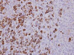
- Immunohistochemical analysis of paraffin-embedded Hs578T xenograft, using XIAP (Product # PA5-29253) antibody at 1:500 dilution. Antigen Retrieval: EDTA based buffer, pH 8.0, 15 min.
Supportive validation
- Submitted by
- Invitrogen Antibodies (provider)
- Main image
- Experimental details
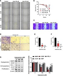
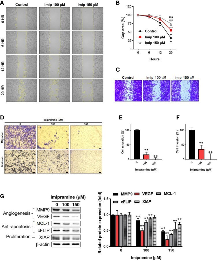
- Figure 4 The suppression of NF-kappaB mediated metastasis ability by imipramine in CL1-5-F4 cells. (A, B) The migration pattern and quantification of gap area after treatment with 100 and 150 muM imipramine are performed by wound healing assay and displayed. (C) The crystal violet staining results of wound healing assay after 20 hr migration are presented. (D-F) The transwell migration, invasion and quantification bar chart after imipramine treatment are displayed. (G) The protein expression of MMP9, VEGF, MCL-1, cFLIP, XIAP and their quantification results are displayed. ( ** p < 0.01 vs. 0 muM imipramine; ## p < 0.01 vs . 100 muM imipramine).
- Submitted by
- Invitrogen Antibodies (provider)
- Main image
- Experimental details
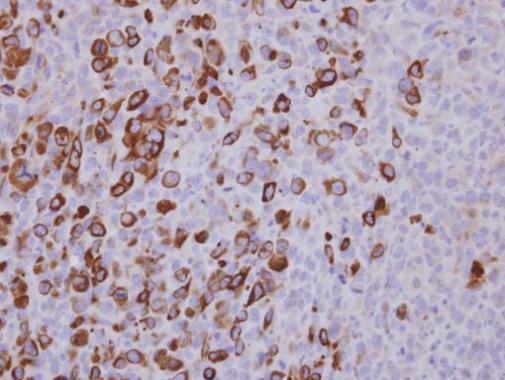
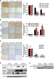
- Figure 6 The inhibition of EGFR/PKC-delta/NF-kappaB proteins phosphorylation and induction of apoptosis-related proteins by imipramine in CL1-5-F4/ NF-kappaB-luc2 bearing tumor. (A-C) The protein expression from IHC of EGFR (Try 1068), PKC-delta (Thr507), NF-kappaB (Ser536), cleaved caspase-3, -8, -9, MMP-9, XIAP, MCL-1 and their quantification bar chart are presented. (D, E) The tumor ex vivo Western blotting from each mice of cleaved caspase-3, -8, -9 and PARP-1 is presented. ( ** p < 0.01 vs . vehicle; scale bar =100 mum).
- Submitted by
- Invitrogen Antibodies (provider)
- Main image
- Experimental details
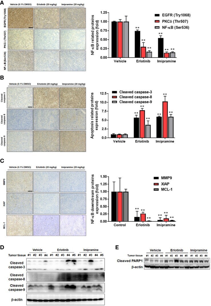
- Figure 1 Effect of magnolol on cell viability and apoptosis in SK-Hep1 cells. Cells were treated with 0-150 muM magnolol for 48 h. A) Evaluationof cell viability was executed by the MTT assay. B) Cell-cycle distribution and C) caspase-3 activity were investigated with flow cytometry. D)Protein levels of survivin and XIAP were evaluated with western blot analysis. *p

- Submitted by
- Invitrogen Antibodies (provider)
- Main image
- Experimental details
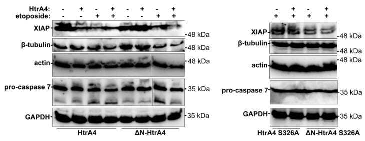
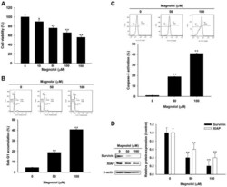
- Figure 9 HtrA4 promotes degradation of XIAP and, less efficiently, of beta-tubulin, actin, and pro-caspase 7. The A549 cells with exogenous HtrA4, DeltaN-HtrA4, or their inactive variants (+) induced by adding doxycycline to the medium were treated with 15 uM etoposide for 48 h. Control cells (-) were incubated with etoposide but without doxycycline. The cells and medium were collected and probed with specific antibodies. Representative blots are presented. Densitometric analyses of the immunoblotting results are shown in Figure S5 .