 Explore
 Validate
 Learn
Learn Western blot
Antibody data
- Antibody Data
- Antigen structure
- References [13]
- Comments [0]
- Validations
- Western blot [3]
- Immunohistochemistry [2]
Submit
Validation data
Reference
Comment
Report error
- Product number
- AF8221 - Provider product page

- Provider
- R&D Systems
- Product name
- Human/Mouse/Rat XIAP Antibody
- Antibody type
- Polyclonal
- Description
- Antigen Affinity-purified. Detects human, mouse and rat XIAP in Western blots.
- Reactivity
- Human, Mouse, Rat
- Host
- Goat
- Conjugate
- Unconjugated
- Antigen sequence
P98170- Isotype
- IgG
- Vial size
- 100 ug
- Concentration
- LYOPH
- Storage
- Use a manual defrost freezer and avoid repeated freeze-thaw cycles. 12 months from date of receipt, -20 to -70 °C as supplied. 1 month, 2 to 8 °C under sterile conditions after reconstitution. 6 months, -20 to -70 °C under sterile conditions after reconstitution.
Submitted references Chronic oxycodone induces axonal degeneration in rat brain.
ASTX660, a Novel Non-peptidomimetic Antagonist of cIAP1/2 and XIAP, Potently Induces TNFα-Dependent Apoptosis in Cancer Cell Lines and Inhibits Tumor Growth.
Delayed apoptosis allows islet β-cells to implement an autophagic mechanism to promote cell survival.
Anticancer efficacy of the hypoxia-activated prodrug evofosfamide is enhanced in combination with proapoptotic receptor agonists against osteosarcoma.
Delivery of a survivin promoter-driven antisense survivin-expressing plasmid DNA as a cancer therapeutic: a proof-of-concept study.
Pro-Apoptotic Activity of New Honokiol/Triphenylmethane Analogues in B-Cell Lymphoid Malignancies.
Admixture fine-mapping in African Americans implicates XAF1 as a possible sarcoidosis risk gene.
Cytotoxic activity of the amphibian ribonucleases onconase and r-amphinase on tumor cells from B cell lymphoproliferative disorders.
Doxorubicin overcomes resistance to drozitumab by antagonizing Inhibitor of Apoptosis Proteins (IAPs).
Serum levels of inhibitors of apoptotic proteins (IAPs) change with IVIg therapy in pemphigus.
Acquired resistance to TRAIL-induced apoptosis in human ovarian cancer cells is conferred by increased turnover of mature caspase-3.
Involvement of the Edar signaling in the control of hair follicle involution (catagen).
Activation of NF-kappaB and upregulation of intracellular anti-apoptotic proteins via the IGF-1/Akt signaling in human multiple myeloma cells: therapeutic implications.
Fan R, Schrott LM, Arnold T, Snelling S, Rao M, Graham D, Cornelius A, Korneeva NL
BMC neuroscience 2018 Mar 23;19(1):15
BMC neuroscience 2018 Mar 23;19(1):15
ASTX660, a Novel Non-peptidomimetic Antagonist of cIAP1/2 and XIAP, Potently Induces TNFα-Dependent Apoptosis in Cancer Cell Lines and Inhibits Tumor Growth.
Ward GA, Lewis EJ, Ahn JS, Johnson CN, Lyons JF, Martins V, Munck JM, Rich SJ, Smyth T, Thompson NT, Williams PA, Wilsher NE, Wallis NG, Chessari G
Molecular cancer therapeutics 2018 Jul;17(7):1381-1391
Molecular cancer therapeutics 2018 Jul;17(7):1381-1391
Delayed apoptosis allows islet β-cells to implement an autophagic mechanism to promote cell survival.
Hayes HL, Peterson BS, Haldeman JM, Newgard CB, Hohmeier HE, Stephens SB
PloS one 2017;12(2):e0172567
PloS one 2017;12(2):e0172567
Anticancer efficacy of the hypoxia-activated prodrug evofosfamide is enhanced in combination with proapoptotic receptor agonists against osteosarcoma.
Liapis V, Zysk A, DeNichilo M, Zinonos I, Hay S, Panagopoulos V, Shoubridge A, Difelice C, Ponomarev V, Ingman W, Atkins GJ, Findlay DM, Zannettino ACW, Evdokiou A
Cancer medicine 2017 Sep;6(9):2164-2176
Cancer medicine 2017 Sep;6(9):2164-2176
Delivery of a survivin promoter-driven antisense survivin-expressing plasmid DNA as a cancer therapeutic: a proof-of-concept study.
Lin KY, Cheng SM, Tsai SL, Tsai JY, Lin CH, Cheung CH
OncoTargets and therapy 2016;9:2601-13
OncoTargets and therapy 2016;9:2601-13
Pro-Apoptotic Activity of New Honokiol/Triphenylmethane Analogues in B-Cell Lymphoid Malignancies.
Mędra A, Witkowska M, Majchrzak A, Cebula-Obrzut B, Bonner MY, Robak T, Arbiser JL, Smolewski P
Molecules (Basel, Switzerland) 2016 Jul 30;21(8)
Molecules (Basel, Switzerland) 2016 Jul 30;21(8)
Admixture fine-mapping in African Americans implicates XAF1 as a possible sarcoidosis risk gene.
Levin AM, Iannuzzi MC, Montgomery CG, Trudeau S, Datta I, Adrianto I, Chitale DA, McKeigue P, Rybicki BA
PloS one 2014;9(3):e92646
PloS one 2014;9(3):e92646
Cytotoxic activity of the amphibian ribonucleases onconase and r-amphinase on tumor cells from B cell lymphoproliferative disorders.
Smolewski P, Witkowska M, Zwolinska M, Cebula-Obrzut B, Majchrzak A, Jeske A, Darzynkiewicz Z, Ardelt W, Ardelt B, Robak T
International journal of oncology 2014 Jul;45(1):419-25
International journal of oncology 2014 Jul;45(1):419-25
Doxorubicin overcomes resistance to drozitumab by antagonizing Inhibitor of Apoptosis Proteins (IAPs).
Zinonos I, Labrinidis A, Liapis V, Hay S, Panagopoulos V, Denichilo M, Ponomarev V, Ingman W, Atkins GJ, Findlay DM, Zannettino AC, Evdokiou A
Anticancer research 2014 Dec;34(12):7007-20
Anticancer research 2014 Dec;34(12):7007-20
Serum levels of inhibitors of apoptotic proteins (IAPs) change with IVIg therapy in pemphigus.
Toosi S, Habib N, Torres G, Reynolds SR, Bystryn JC
The Journal of investigative dermatology 2011 Nov;131(11):2327-9
The Journal of investigative dermatology 2011 Nov;131(11):2327-9
Acquired resistance to TRAIL-induced apoptosis in human ovarian cancer cells is conferred by increased turnover of mature caspase-3.
Lane D, Côté M, Grondin R, Couture MC, Piché A
Molecular cancer therapeutics 2006 Mar;5(3):509-21
Molecular cancer therapeutics 2006 Mar;5(3):509-21
Involvement of the Edar signaling in the control of hair follicle involution (catagen).
Fessing MY, Sharova TY, Sharov AA, Atoyan R, Botchkarev VA
The American journal of pathology 2006 Dec;169(6):2075-84
The American journal of pathology 2006 Dec;169(6):2075-84
Activation of NF-kappaB and upregulation of intracellular anti-apoptotic proteins via the IGF-1/Akt signaling in human multiple myeloma cells: therapeutic implications.
Mitsiades CS, Mitsiades N, Poulaki V, Schlossman R, Akiyama M, Chauhan D, Hideshima T, Treon SP, Munshi NC, Richardson PG, Anderson KC
Oncogene 2002 Aug 22;21(37):5673-83
Oncogene 2002 Aug 22;21(37):5673-83
No comments: Submit comment
Supportive validation
- Submitted by
- R&D Systems (provider)
- Main image
- Experimental details
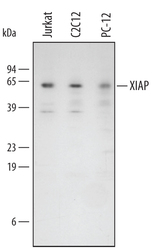
- Detection of Human/Mouse/Rat XIAP by Western Blot. Western blot shows lysates of Jurkat human acute T cell leukemia cell line, C2C12 mouse myoblast cell line, and PC-12 rat adrenal pheochromocytoma cell line. PVDF membrane was probed with 0.5 µg/mL of Goat Anti-Human/Mouse/Rat XIAP Antigen Affinity-purified Polyclonal Antibody (Catalog # AF8221) followed by HRP-conjugated Anti-Goat IgG Secondary Antibody (Catalog # HAF109). A specific band was detected for XIAP at approximately 56 kDa (as indicated). This experiment was conducted under reducing conditions and using Immunoblot Buffer Group 4.
- Submitted by
- R&D Systems (provider)
- Main image
- Experimental details
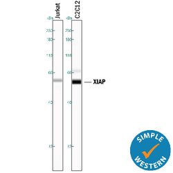
- Detection of Human and Mouse XIAP by Simple WesternTM. Simple Western lane view shows lysates of Jurkat human acute T cell leukemia cell line and C2C12 mouse myoblast cell line, loaded at 0.2 mg/mL. A specific band was detected for XIAP at approximately 59 kDa (as indicated) using 5 µg/mL of Goat Anti-Human/Mouse/Rat XIAP Antigen Affinity-purified Polyclonal Antibody (Catalog # AF8221) followed by 1:50 dilution of HRP-conjugated Anti-Goat IgG Secondary Antibody (Catalog # HAF109). This experiment was conducted under reducing conditions and using the 12-230 kDa separation system.

- Submitted by
- R&D Systems (provider)
- Main image

- Experimental details
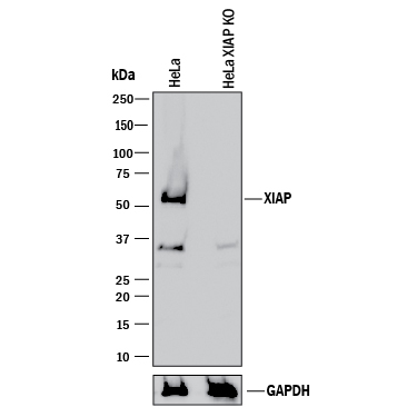
- Western Blot Shows Human XIAP Specificity by Using Knockout Cell Line. Western blot shows lysates of HeLa human cervical epithelial carcinoma parental cell line and XIAP knockout HeLa cell line (KO). PVDF membrane was probed with 0.5 µg/mL of Goat Anti-Human/Mouse/Rat XIAP Antigen Affinity-purified Polyclonal Antibody (Catalog # AF8221) followed by HRP-conjugated Anti-Goat IgG Secondary Antibody (Catalog # HAF017). A specific band was detected for XIAP at approximately 52 kDa (as indicated) in the parental HeLa cell line, but is not detectable in knockout HeLa cell line. GAPDH (Catalog # AF5718) is shown as a loading control. This experiment was conducted under reducing conditions and using Immunoblot Buffer Group 1.
Supportive validation
- Submitted by
- R&D Systems (provider)
- Main image
- Experimental details
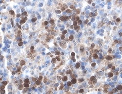
- XIAP in Human Lymphoma. XIAP was detected in immersion fixed paraffin-embedded sections of human lymphoma using 5 µg/mL Goat Anti-Human/Mouse/Rat XIAP Antigen Affinity-purified Polyclonal Antibody (Catalog # AF8221) overnight at 4 °C. Tissue was stained with the Anti-Goat HRP-DAB Cell & Tissue Staining Kit (brown; Catalog # CTS008) and counterstained with hematoxylin (blue). View our protocol for Chromogenic IHC Staining of Paraffin-embedded Tissue Sections.

- Submitted by
- R&D Systems (provider)
- Main image
- Experimental details
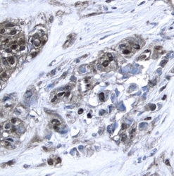
- XIAP in Human Breast Cancer Tissue. XIAP was detected in immersion fixed paraffin-embedded sections of human breast cancer tissue using 15 µg/mL Goat Anti-Human/Mouse/Rat XIAP Antigen Affinity-purified Polyclonal Antibody (Catalog # AF8221) overnight at 4 °C. Tissue was stained with the Anti-Goat HRP-DAB Cell & Tissue Staining Kit (brown; Catalog # CTS008) and counterstained with hematoxylin (blue). View our protocol for Chromogenic IHC Staining of Paraffin-embedded Tissue Sections.
