Explore
 Validate
 Learn
Learn Immunocytochemistry
Antibody data
- Antibody Data
- Antigen structure
- References [1]
- Comments [0]
- Validations
- Immunocytochemistry [1]
- Immunohistochemistry [1]
Submit
Validation data
Reference
Comment
Report error
- Product number
- HPA028888 - Provider product page

- Provider
- Atlas Antibodies
- Proper citation
- Atlas Antibodies Cat#HPA028888, RRID:AB_2672814
- Product name
- Anti-DAB2
- Antibody type
- Polyclonal
- Description
- Polyclonal Antibody against Human DAB2, Gene description: Dab, mitogen-responsive phosphoprotein, homolog 2 (Drosophila), Alternative Gene Names: DOC-2, Validated applications: ICC, IHC, Uniprot ID: P98082, Storage: Store at +4°C for short term storage. Long time storage is recommended at -20°C.
- Reactivity
- Human
- Host
- Rabbit
- Conjugate
- Unconjugated
- Isotype
- IgG
- Vial size
- 100 µl
- Concentration
- 0.2 mg/ml
- Storage
- Store at +4°C for short term storage. Long time storage is recommended at -20°C.
- Handling
- The antibody solution should be gently mixed before use.
Submitted references Highly efficient generation of self-renewing trophoblast from human pluripotent stem cells.
Slamecka J, Ryu S, Tristan CA, Chu PH, Weber C, Deng T, Gedik Y, Ormanoglu P, Voss TC, Simeonov A, Singeç I
iScience 2024 Oct 18;27(10):110874
iScience 2024 Oct 18;27(10):110874
No comments: Submit comment
Supportive validation
- Submitted by
- Atlas Antibodies (provider)
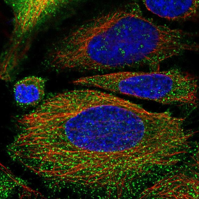
- Main image

- Experimental details
- Immunofluorescent staining of human cell line HeLa shows localization to nucleoli fibrillar center, plasma membrane & vesicles.
- Sample type
- Human
Supportive validation
- Submitted by
- Atlas Antibodies (provider)
- Enhanced method
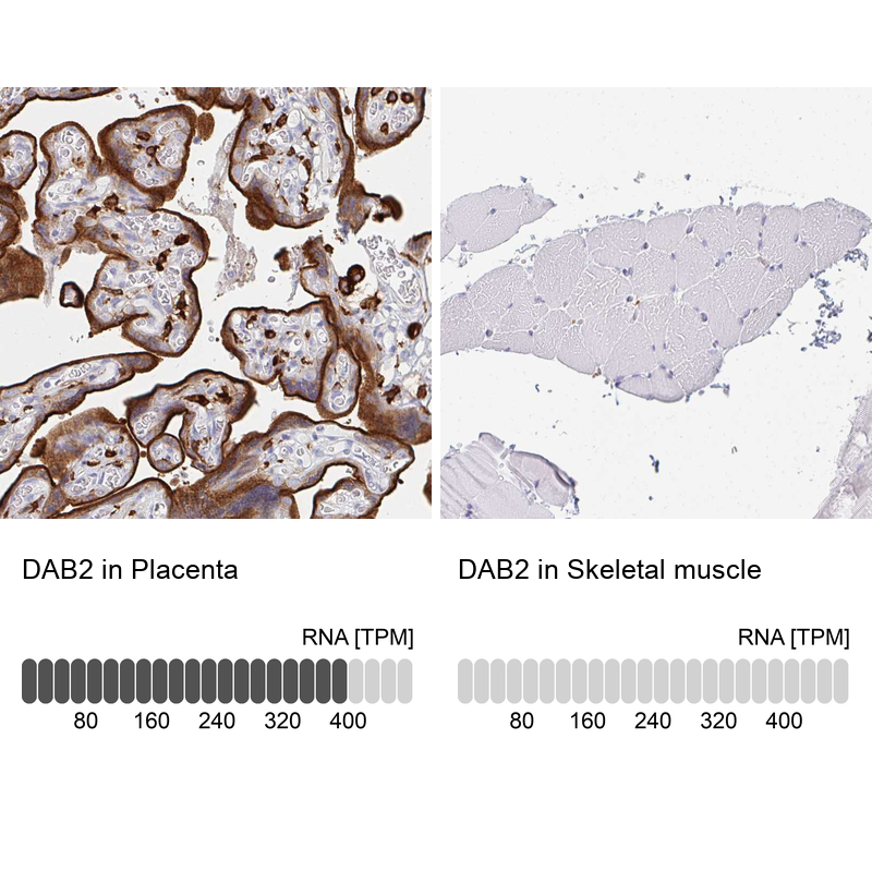
- Orthogonal validation
- Main image

- Experimental details
- Immunohistochemistry analysis in human placenta and skeletal muscle tissues using HPA028888 antibody. Corresponding DAB2 RNA-seq data are presented for the same tissues.
- Sample type
- Human
- Protocol
- Protocol