 Explore
 Validate
 Learn
Learn Western blot
Antibody data
- Antibody Data
- Antigen structure
- References [3]
- Comments [0]
- Validations
- Western blot [1]
- Immunocytochemistry [1]
- Flow cytometry [1]
Submit
Validation data
Reference
Comment
Report error
- Product number
- MA1-189 - Provider product page

- Provider
- Invitrogen Antibodies
- Product name
- TBP Monoclonal Antibody (1TBP18)
- Antibody type
- Monoclonal
- Antigen
- Other
- Description
- MA1-189 detects TATA-Binding Protein from human, mouse, and rat, canine, and non-human primate samples. TBP (human) has an approximate molecular weight of 38kDa, while other species' isoforms can range from 35-36kDa. MA1-189 has been successfully used in Western blot and immunofluorescence and flow cytometry applications. The MA1-189 immunogen is the N-terminal domain of the human TATA-binding protein (TBP).
- Reactivity
- Human, Mouse, Rat, Canine
- Host
- Mouse
- Isotype
- IgG
- Antibody clone number
- 1TBP18
- Vial size
- 100 µL
- Concentration
- 1 mg/mL
- Storage
- -20° C, Avoid Freeze/Thaw Cycles
Submitted references FAIM-S functions as a negative regulator of NF-κB pathway and blocks cell cycle progression in NSCLC cells.
Modeling the Function of TATA Box Binding Protein in Transcriptional Changes Induced by HIV-1 Tat in Innate Immune Cells and the Effect of Methamphetamine Exposure.
A non-canonical BRD9-containing BAF chromatin remodeling complex regulates naive pluripotency in mouse embryonic stem cells.
Wang P, Xun W, Han T, Cheng Z
Cell cycle (Georgetown, Tex.) 2020 Dec;19(24):3458-3467
Cell cycle (Georgetown, Tex.) 2020 Dec;19(24):3458-3467
Modeling the Function of TATA Box Binding Protein in Transcriptional Changes Induced by HIV-1 Tat in Innate Immune Cells and the Effect of Methamphetamine Exposure.
Tjitro R, Campbell LA, Basova L, Johnson J, Najera JA, Lindsey A, Marcondes MCG
Frontiers in immunology 2018;9:3110
Frontiers in immunology 2018;9:3110
A non-canonical BRD9-containing BAF chromatin remodeling complex regulates naive pluripotency in mouse embryonic stem cells.
Gatchalian J, Malik S, Ho J, Lee DS, Kelso TWR, Shokhirev MN, Dixon JR, Hargreaves DC
Nature communications 2018 Dec 3;9(1):5139
Nature communications 2018 Dec 3;9(1):5139
No comments: Submit comment
Supportive validation
- Submitted by
- Invitrogen Antibodies (provider)
- Main image

- Experimental details
- Western blot analysis of TBP was performed by loading 20 µg of the indicated whole cell lysates and 5 µL of PageRuler Plus Prestained Protein Ladder (Product # 26619) per well onto a Novex 4-20% Tris-Glycine polyacrylamide gel (Product # WT4202BOX ). Proteins were transferred to a nitrocellulose membrane using the G2 Blotter (Product # 62288), and blocked with 5% Milk in TBST for 1 hour at room temperature. TATA binding protein was detected at ~36-38 kD using a TBP monoclonal antibody (Product # MA1-189) at a dilution of 1:1000 in 5% Milk in TBST overnight at 4C on a rocking platform, followed by a Goat anti-Mouse IgG (H+L) Superclonal Secondary Antibody, HRP conjugate (Product # A28177) at a dilution of 1:1000 for at least 30 minutes at room temperature. Chemiluminescent detection was performed using SuperSignal Pico substrate (Product # 34078) and the myECL Imager (Product # 62236).

Supportive validation
- Submitted by
- Invitrogen Antibodies (provider)
- Main image
- Experimental details
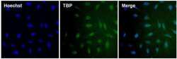
- Immunofluorescent analysis of TATA binding protein (green) in U2OS cells. The cells were fixed with 4% paraformaldehyde for 15 minutes, permeabilized with 0.1% Triton X-100 in PBS for 15 minutes, and blocked with 3% BSA in PBS (Product # 37525) for 30 minutes at room temperature. Cells were stained with a TBP monoclonal antibody (Product # MA1-189) at a dilution of 10 µg/mL in staining buffer for 1 hour at room temperature, and then incubated with a Goat anti-Mouse IgG Superclonal Secondary Antibody, Alexa Fluor 488 conjugate (Product # A28175) at a dilution of 1:250 for 1 hour at room temperature (green). Nuclei (blue) were counterstained with Hoechst 33342 dye (Product # 62249). Images were taken on a Thermo Scientific ToxInsight Instrument at 20X magnification.

Supportive validation
- Submitted by
- Invitrogen Antibodies (provider)
- Main image

- Experimental details
- Flow cytometry analysis of TATA binding protein was done on HeLa cells. Cells were fixed, permeabilized and stained with a TBP mouse monoclonal antibody (Product # MA1-189, pink histogram) or an isotype control (Product # MA1-10406, black histogram) at a dilution of 5 µg/mL. After incubation for 1 hour on ice, the cells were labeled with DyLight 650 Goat Anti-Mouse Secondary Antibody (Product # 84545) at a dilution of 1:50 for 1 hour on ice. A representative 10,000 cells were acquired and analyzed for each sample.
