Explore
 Validate
 Learn
Learn Western blot
Antibody data
- Antibody Data
- Antigen structure
- References [3]
- Comments [0]
- Validations
- Western blot [1]
- Immunohistochemistry [1]
Submit
Validation data
Reference
Comment
Report error
- Product number
- MAB5175 - Provider product page

- Provider
- R&D Systems
- Product name
- Mouse Uromodulin Antibody
- Antibody type
- Monoclonal
- Description
- Protein A or G purified from hybridoma culture supernatant. Detects mouse Uromodulin in direct ELISAs and Western blots. In direct ELISAs, 100% cross-reactivity with recombinant human Uromodulin is observed.
- Reactivity
- Mouse
- Host
- Rat
- Conjugate
- Unconjugated
- Antigen sequence
Q91X17- Isotype
- IgG
- Antibody clone number
- 774056
- Vial size
- 100 ug
- Concentration
- LYOPH
- Storage
- Use a manual defrost freezer and avoid repeated freeze-thaw cycles. 12 months from date of receipt, -20 to -70 °C as supplied. 1 month, 2 to 8 °C under sterile conditions after reconstitution. 6 months, -20 to -70 °C under sterile conditions after reconstitution.
Submitted references Repression of Interstitial Identity in Nephron Progenitor Cells by Pax2 Establishes the Nephron-Interstitium Boundary during Kidney Development.
Missense Mutation of POU Domain Class 3 Transcription Factor 3 in Pou3f3L423P Mice Causes Reduced Nephron Number and Impaired Development of the Thick Ascending Limb of the Loop of Henle.
Botulinum toxin A complex exploits intestinal M cells to enter the host and exert neurotoxicity.
Naiman N, Fujioka K, Fujino M, Valerius MT, Potter SS, McMahon AP, Kobayashi A
Developmental cell 2017 May 22;41(4):349-365.e3
Developmental cell 2017 May 22;41(4):349-365.e3
Missense Mutation of POU Domain Class 3 Transcription Factor 3 in Pou3f3L423P Mice Causes Reduced Nephron Number and Impaired Development of the Thick Ascending Limb of the Loop of Henle.
Rieger A, Kemter E, Kumar S, Popper B, Aigner B, Wolf E, Wanke R, Blutke A
PloS one 2016;11(7):e0158977
PloS one 2016;11(7):e0158977
Botulinum toxin A complex exploits intestinal M cells to enter the host and exert neurotoxicity.
Matsumura T, Sugawara Y, Yutani M, Amatsu S, Yagita H, Kohda T, Fukuoka S, Nakamura Y, Fukuda S, Hase K, Ohno H, Fujinaga Y
Nature communications 2015 Feb 17;6:6255
Nature communications 2015 Feb 17;6:6255
No comments: Submit comment
Supportive validation
- Submitted by
- R&D Systems (provider)
- Main image
- Experimental details
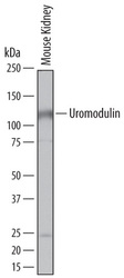
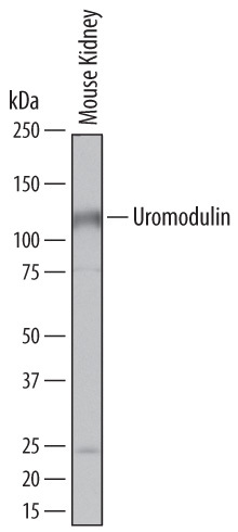
- Detection of Mouse Uromodulin by Western Blot. Western blot shows lysates of mouse kidney tissue. PVDF membrane was probed with 0.5 µg/mL of Rat Anti-Mouse Uromodulin Monoclonal Antibody (Catalog # MAB5175) followed by HRP-conjugated Anti-Rat IgG Secondary Antibody (Catalog # HAF005). A specific band was detected for Uromodulin at approximately 115 kDa (as indicated). This experiment was conducted under reducing conditions and using Immunoblot Buffer Group 8.
Supportive validation
- Submitted by
- R&D Systems (provider)
- Main image
- Experimental details
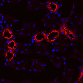
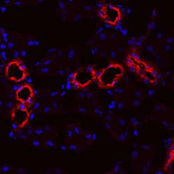
- Uromodulin in Mouse Kidney. Uromodulin was detected in immersion fixed frozen sections of mouse kidney using Rat Anti-Mouse Uromodulin Monoclonal Antibody (Catalog # MAB5175) at 10 µg/mL overnight at 4 °C. Tissue was stained using the NorthernLights™ 557-conjugated Anti-Rat IgG Secondary Antibody (red; Catalog # NL013) and counterstained with DAPI (blue). Specific staining was localized to convoluted tubule epithelial cells. View our protocol for Fluorescent IHC Staining of Frozen Tissue Sections.