 Explore
 Validate
 Learn
Learn Western blot
Antibody data
- Antibody Data
- Antigen structure
- References [2]
- Comments [0]
- Validations
- Western blot [3]
- Chromatin Immunoprecipitation [1]
- Other assay [1]
Submit
Validation data
Reference
Comment
Report error
- Product number
- PA1-882 - Provider product page

- Provider
- Invitrogen Antibodies
- Product name
- DNMT3A Polyclonal Antibody
- Antibody type
- Polyclonal
- Antigen
- Synthetic peptide
- Description
- PA1-882 detects DNA methyltransferase 3a (Dnmt3a) from human and mouse tissues and cells as well as recombinant human Dnmt3a. This antibody does not detect full length recombinant human Dnmt3b or Dnmt1. PA1-882 has been successfully used in Western blot procedures. By Western blot, this antibody detects an ~120 kDa protein representing Dnmt3a from P19 cell nuclear extracts. The PA1-882 immunogen is a synthetic peptide corresponding to residues D(86) L L P N G D L E K R S E P Q(100) C of human Dnmt3a. This immunizing peptide (Cat. # PEP-116) is available for use in neutralization and control experiments.
- Reactivity
- Human, Mouse
- Host
- Rabbit
- Isotype
- IgG
- Vial size
- 100 µg
- Concentration
- 1 mg/mL
- Storage
- -20° C, Avoid Freeze/Thaw Cycles
Submitted references Sex-specific lung functional changes in adult mice exposed only to second-hand smoke in utero.
Preferential methylation of unmethylated DNA by Mammalian de novo DNA methyltransferase Dnmt3a.
Noël A, Xiao R, Perveen Z, Zaman H, Le Donne V, Penn A
Respiratory research 2017 Jun 27;18(1):104
Respiratory research 2017 Jun 27;18(1):104
Preferential methylation of unmethylated DNA by Mammalian de novo DNA methyltransferase Dnmt3a.
Yokochi T, Robertson KD
The Journal of biological chemistry 2002 Apr 5;277(14):11735-45
The Journal of biological chemistry 2002 Apr 5;277(14):11735-45
No comments: Submit comment
Supportive validation
- Submitted by
- Invitrogen Antibodies (provider)
- Main image

- Experimental details
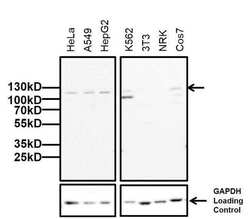
- Western blot analysis of Dnmt3a was performed by loading 20 µg of the indicated whole cell lysates and 5 µL of PageRuler Plus Prestained Protein Ladder (Product # 26619) onto a 4-20% Tris-Glycine polyacrylamide gel (Product # WT4202BX10). Proteins were transferred to a nitrocellulose membrane using the G2 Blotter (Product # 62288), and blocked with 5% Milk in TBST for 1 hour at room temperature. Dnmt3a was detected at ~120 kDa using a Dnmt3a rabbit polyclonal antibody (Product # PA1-882) at a dilution of 1:500 in blocking buffer overnight at 4C on a rocking platform, followed by a Goat anti-rabbit IgG (H+L) Superclonal™ Secondary Antibody, HRP conjugate (Product # A27036) at a dilution of 1:1000 for at least one hour at room temperature. Chemiluminescent detection was performed using SuperSignal West Pico (Product # 34078).

- Submitted by
- Invitrogen Antibodies (provider)
- Main image
- Experimental details
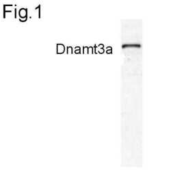
- Western blot of Dnmt3a on P19 cell nuclear extract using Product # PA1-882.

- Submitted by
- Invitrogen Antibodies (provider)
- Main image
- Experimental details
- Western blot analysis of Dnmt3a was performed by loading 20 µg of the indicated whole cell lysates and 5 µL of PageRuler Plus Prestained Protein Ladder (Product # 26619) onto a 4-20% Tris-Glycine polyacrylamide gel (Product # WT4202BX10). Proteins were transferred to a nitrocellulose membrane using the G2 Blotter (Product # 62288), and blocked with 5% Milk in TBST for 1 hour at room temperature. Dnmt3a was detected at ~120 kDa using a Dnmt3a rabbit polyclonal antibody (Product # PA1-882) at a dilution of 1:1000 in blocking buffer overnight at 4C on a rocking platform, followed by a Goat anti-rabbit IgG (H+L) Superclonal™ Secondary Antibody, HRP conjugate (Product # A27036) at a dilution of 1:2000 for at least one hour at room temperature. Chemiluminescent detection was performed using SuperSignal West Pico (Product # 34078).

Supportive validation
- Submitted by
- Invitrogen Antibodies (provider)
- Main image

- Experimental details
- Multiplex microplate Matrix ChIP has been described in detail (http://www.ncbi.nlm.nih.gov/pubmed/25959381). Briefly HTC116 cells were starved followed by addition of serum and samples of cells were cross-linked with formaldehyde after the time points indicated on the x-axis (0, 5, 15 and 30 min). Chromatin was sheared using a Bioruptor and ChIP assays were performed using protein A-coated 96-well polypropylene microplates with 1uL/100uL well volume of Dnmt3a polyclonal antibody (Product # PA1-882). Quantitative real-time PCRs were performed in quadruplicate using 1 to 2uL of DNA with primers to -15kb downstream of Egr1 exon 1 of Erg1 and exon 2 of Erg1. PCR calibration curves were generated for each primer pair from a dilution series of total human genomic DNA. The PCR primer efficiency curve was fit to cycle threshold (Ct) versus log [genomic DNA concentration] by using an r2 best fit. DNA concentration values for each ChIP and input DNA sample were calculated from their respective average Ct values. Final results are expressed as fraction of input DNA. Schematic representations of the Erg1 loci is shown where boxes represent exons (black boxes = translated regions, white boxes = untranslated regions), the zigzag line represents an intron, and the straight line represents upstream sequence. Regions amplified by the primers are represented by black bars. Data courtesy of Dr. Karol Bomsztyk’s laboratory.

Supportive validation
- Submitted by
- Invitrogen Antibodies (provider)
- Main image

- Experimental details
- Fig. 5 In utero SHS dysregulates the protein expression of DNMT3A, SERPINA1A, MAPK7, and PHF1 in male and female mice. a ) Western blots show that DNMT3A and SERPINA1A were down-regulated, whereas MAPK7 and PHF1 were up-regulated in BALB/c male mice exposed in utero to SHS versus air-treated controls. b ) Western blots show that SERPINA1A was down-regulated, whereas DNMT3A and MAPK7 were up-regulated in BALB/c female mice exposed in utero to SHS versus air-treated controls. c ) Mean densitometry +- SEM results showing fold change of treated mice versus controls for the proteins analyzed
